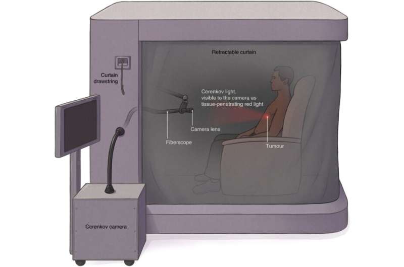
A team of researchers affiliated with several institutions in the U.S. and one in the U.K. has developed a new type of Cerenkov luminescence imaging device to help doctors spot cancerous tumors. In their paper published in the journal Nature Biomedical Engineering, the group describes the device, how it works and possible uses for it in clinical settings.
Cerenkov luminescence is a type of electromagnetic radiation where charged particles pass through a dielectric medium at a speed that is faster than regular light in the medium. It is commonly measured by astrophysicists looking at stars and other researchers working in nuclear reactors. Prior research has shown that Cerenkov luminescence can also be generated in the human body as light passes through tissue. It has been suggested that Cerenkov luminescence could be used to help differentiate tumor tissue from normal tissue, and some work has been done to create Cerenkov luminescence imaging (CLI) devices. But to date, such devices have suffered from a variety of issues that have prevented their use in clinical settings. In this new effort, the researchers have designed and built a complete CLI device that has already passed an initial clinical trial.
One of the problems with CLI devices in the past was interference by ambient light—to overcome that problem, use of the new device involves having a patient sit inside a completely enclosed chamber that blocks all other light sources. Inside, CLI particles are released via radiotracers that result in target tissue vibrating in a way that releases light that can be captured by a camera.
Source: Read Full Article
