Severe acute respiratory syndrome coronavirus 2 (SARS-CoV-2) infection incites adaptive cellular immune responses. In many SARS-CoV-2 studies, peripheral blood is analyzed to study the immune responses. Now, a new study under consideration at the Nature Portfolio Journal analyzes the peripheral blood, tonsils, and adenoids of children to understand local and systemic immune responses to SARS-CoV-2. A preprint version of the research paper is available at Research Square, while it undergoes peer review.
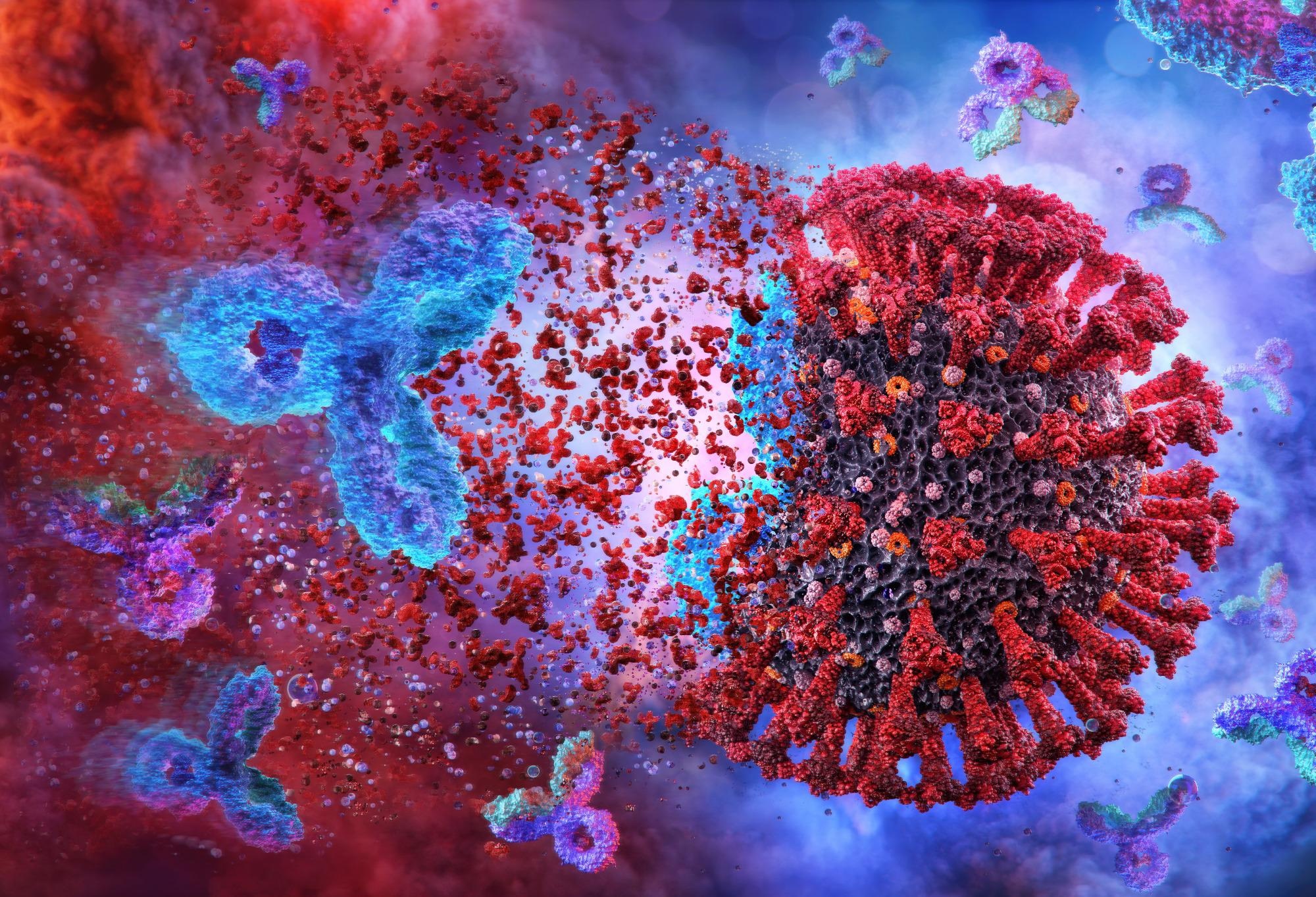
 Study: Robust, persistent adaptive immune responses to SARS-CoV-2 in the oropharyngeal lymphoid tissue of children. Image Credit: Corona Borealis Studio / Shutterstock
Local adaptive immune responses
SARS-CoV-2 infection and replication occur in the upper respiratory tract. The lymph glands closest to the site of entry of the virus are tonsils and adenoids, present in the nose and throat area. Here, tissue-specific T- and B-cell responses are generated against SARS-CoV-2 antigens in the upper respiratory tract. Tonsillectomy and adenoidectomy are common surgeries in children. Tonsils and adenoids allow the study of local adaptive immune responses.
Cellular interactions in the lymph glands
The activation and maturation of the lymphatic T and B cells occur in the lymph glands. The T follicular helper cells (Tfh) and B cells interact collaboratively to allow immunoglobulin gene class switching. This promotes the formation of germinal centers (GCs) where B cells mature, resulting in the production of antibodies and memory B cells.
Studies have shown that in adults with fatal coronavirus disease 2019 (COVID-19), the serum antibody levels are short-lived because of loss of GCs from thoracic lymph nodes. Conversely, some studies have shown enduring B cell immunity derived from GCs. In addition, some studies have also demonstrated the presence of Tfh cells in the lymph glands and lung tissues organ donors.
GC responses in tissues from lymph glands
In this study, the investigators collected peripheral blood, tonsils, and adenoids from 110 children undergoing tonsillectomy or adenoidectomy.
All participants were COVID-19 negative as tested by an RT-PCR test 72 hours before the surgery. Twenty-four participants showed evidence of a previous SARS-CoV-2 infection, with a confirmed positive RT-PCR test or presence of neutralizing antibodies in the serum.
Neutralizing antibodies against the early SARS-CoV-2 strain WA-1 and six other variants of interest, viz. epsilon, alpha, gamma, beta, iota, delta, were observed in most COVID-19 convalescent subjects. Only 9 out of 23 participants had neutralizing antibodies against the omicron variant.
Neutralizing antibody levels were highest against the WA-1 strain, and the levels reduced with time since the SARS-CoV-2 infection. Except for two participants, all had SARS-CoV-2-specific B cells in their peripheral blood, tonsils, and adenoids. These S+RBD+ B cells bound specifically to the spike protein S1 domain and receptor-binding domain (RBD).
High-dimensional flow cytometry of the B cell populations showed that the S1+RBD+ B cells were memory B cells. Thus, a memory B cell response was elicited and maintained in the upper respiratory tract. Furthermore, this response was robust since it was observed as long as 10 months post-infection.
Flow cytometry data also showed a considerable portion of GC B cells among the S1+RBD+ B cells in tonsils and adenoids. The GC structures were also confirmed by multiplex immunofluorescence microscopy.
Single-cell analysis of B cells
In tissues from two participants and an uninfected control, the S1+ and S1- B cells were sorted and characterized using CITE-seq (Cellular Indexing of Transcriptomes and Epitopes by Sequencing). This measured the expression of B cell surface markers and sequenced the transcriptome and B cell receptors of single B cells. Results of these experiments showed that a portion of S1+ B cell clones was present in both the tonsils and adenoids. The SARS-CoV-2-specific clones underwent class-switching and somatic hypermutation in GCs.
Post-COVID-19 B and T cell populations in the lymph tissues
The tonsils and adenoids of COVID-19-convalescent children had a lower proportion of naive B and T cells. Particularly in the adenoids, there was an expansion of GC B cell populations. These changes prolonged months after SARS-CoV-2 infection.
The tonsils and adenoids had a higher proportion of GC-Tfh cells and T follicular regulatory (Tfr) cells. GC-Tfh cells signal the B cells to form and maintain the GCs. Moreover, these cells had a phenotype characteristic of tissue-resident memory T cells and were present within the GC. Furthermore, their frequency was directly proportional to the frequency of GC B cells. T cell polyfunctionality evaluation using SPICE (Simplified Presentation of Incredibly Complex Evaluations) showed that the Tfh cells produced cytokines that enable GC formation and B cell antibody secretion. IFN-γ-type response was particularly enriched in the adenoids. These data indicate that these T cells help form and maintain SARS-CoV-2-specific GC responses.
Activated and cytotoxic T cells with increased cytokine production and GC localization were also enriched in the lymph tissues.
Post-COVID-19 T cell populations in the blood
There was an enrichment of activated Tfh cells and stem cell-like memory T cells in the peripheral blood post-COVID-19. Blood samples also had SARS-CoV-2-reactive T cells which were not observed in the lymph glands. These cells were primarily memory cells.
Activated and cytotoxic T cells with increased cytokine production and GC localization were not enriched in the peripheral blood.
Viral RNA in the lymph tissues
RNA isolated from formalin-fixed, paraffin-embedded tonsil and adenoid samples and analyzed using digital droplet PCR showed evidence of SARS-CoV-2 nucleocapsid RNA in multiple samples of COVID-19-convalescent tissues.
The viral copies were present even when the nasal swabs were negative for SARS-CoV-2. Moreover, viral RNA copies correlated with the proportion of S1+RBD+ cells among GC B cells in the tonsils. There was no viral protein detected in any of the samples.
Limitations of the study
In this study, the investigators had no information about the date of SARS-CoV-2 infection. Several participants lacked awareness of having COVID-19. The investigators did not have longitudinal samples to map the duration of immune changes. The antigen-specific T cells in the lymph tissues could not be delineated. The COVID-19-convalescent children underwent tonsillectomy for sleep-disordered breathing. This may be an immunologic disorder and may influence the immune responses to SARS-CoV-2 infection.
Conclusion
This study presents evidence of persistent, localized immunity to SARS-CoV-2. In addition, COVID-19-convalescent children showed robust, lymph-tissue-specific adaptive immune responses weeks to months after acute infection.
*Important notice
Research Square publishes preliminary scientific reports that are not peer-reviewed and, therefore, should not be regarded as conclusive, guide clinical practice/health-related behavior, or treated as established information.
- Kalpana Manthiram, Qin Xu, Pedro Milanez-Almeida et al. (2022) Robust, persistent adaptive immune responses to SARS-CoV-2 in the oropharyngeal lymphoid tissue of children. PREPRINT (Version 1) available at Research Square. https://doi.org/10.21203/rs.3.rs-1276578/v1
Posted in: Medical Research News | Disease/Infection News
Tags: Adenoidectomy, Antibodies, Antibody, Antigen, B Cell, Blood, Breathing, Cell, Children, Coronavirus, Coronavirus Disease COVID-19, covid-19, Cytokine, Cytokines, Cytometry, Flow Cytometry, Frequency, Gene, immunity, Immunoglobulin, Lymph Nodes, Microscopy, Omicron, Phenotype, Protein, Receptor, Research, Respiratory, RNA, SARS, SARS-CoV-2, Severe Acute Respiratory, Severe Acute Respiratory Syndrome, Sleep, Spice, Spike Protein, Surgery, Syndrome, Throat, Tonsil, Tonsillectomy, Virus

Written by
Dr. Shital Sarah Ahaley
Dr. Shital Sarah Ahaley is a medical writer. She completed her Bachelor's and Master's degree in Microbiology at the University of Pune. She then completed her Ph.D. at the Indian Institute of Science, Bengaluru where she studied muscle development and muscle diseases. After her Ph.D., she worked at the Indian Institute of Science, Education, and Research, Pune as a post-doctoral fellow. She then acquired and executed an independent grant from the DBT-Wellcome Trust India Alliance as an Early Career Fellow. Her work focused on RNA binding proteins and Hedgehog signaling.
Source: Read Full Article
