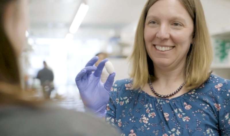
From ski slopes to Girl Scouts, Rosalyn Abbott’s classroom of choice has evolved over the years, but her love for teaching and discovery remains a steady constant. On any given day, she can be found integrating biomaterials, namely silk, with tissue engineering techniques in her lab, or teaching introductory engineering courses to undergraduate students at Carnegie Mellon University. Her group’s latest research uncovered a novel finding—that silk scaffolding is responsive to ultrasound.
Silk is a safe, unique, and versatile natural biomaterial that has successfully been used in wound healing and in tissue engineering of cartilage, tendon, and ligament tissues. It can easily be extracted from several sources, most commonly a Bombyx mori cocoon, and from there, processed into different formats including gels, scaffolds, and films.
“We call silk a ‘blank slate’ because it has the potential to be used in so many different applications,” explained Abbott, an assistant professor of biomedical engineering. “Not only can you alter silk’s mechanical properties, but also, you can adjust its degradation rate, or in other words, how quickly or slowly silk scaffolding material breaks down in the body as it is replaced by new tissue.”
Abbott’s lab uses silk to engineer adipose (fat) tissue depots for filling soft tissue defects and for modeling diseases. This work fits into the bigger picture field of tissue engineering, or the practice of combining scaffolds, cells, and biologically active molecules into functional tissues. Ultimately, the aim is to assemble functional constructs that restore, maintain, or improve damaged tissues or whole organs.
Currently, there is no method of determining a patient’s regenerative rate prior to implanting a biomaterial, nor is there a way to adjust the degradation once the material has been implanted. Patients regenerate tissues at different rates, depending on such factors as age, nutritional status, disease state, lifestyle, gender, etc. A mismatch in regenerative rate and biomaterial degradation leads to inappropriate immune responses and poor healing of the implant.
In recent work published in Advanced Healthcare Materials, Abbott’s group investigated the use of non-invasive, therapeutic ultrasound to trigger and adjust silk scaffold degradation post-implantation. They showed that ultrasound exposure could successfully decrease weight, while increasing porosity of silk scaffolds—without affecting the health or metabolism of cells within the tissues. They also tracked scaffold degradation with imaging ultrasound as a proof-of-concept that a clinician could monitor the degradation and adjust the scaffold properties, as needed, on-demand.
These findings are clinically relevant for tissue engineering approaches that implant silk biomaterials to restore structure, while tissues slowly grow into the space and replace the scaffold material.
Next, Abbott’s group plans to research and test a broader range of degradation profiles to ultimately create more responsive biomaterials. Megan DeBari, lead author of the recent Advanced Healthcare Materials paper and an MSE Ph.D. student in Abbott’s lab, is pursuing a startup company to further develop the approach, through an Innovation Fellowship with Carnegie Mellon’s Swartz Center for Entrepreneurship.
Long-term, Abbott is working toward the development of a personalized biomaterial platform, where biomaterial degradation can be adjusted with focused ultrasound following routine monitoring of how a patient is healing.
Source: Read Full Article
