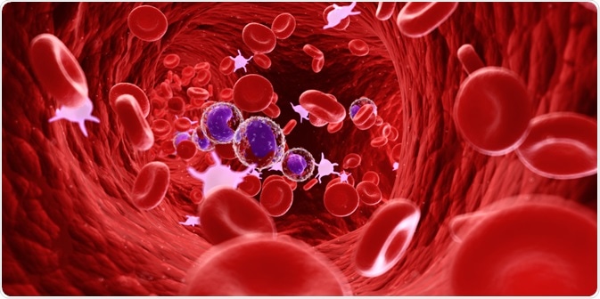
Flow cytometry is a useful technique for the analysis of large populations of cells. This article describes the combination of flow cytometry with other techniques (such as cell sorting) and the applications of these techniques in medicine and research.
This article will cover:
- Fluorescence-activated cell sorting (FACS)
- Coulter counters
- Cytometric bead arrays
- Applications of flow cytometry in medicine and research
 Sebastian Kaulitzki | Shutterstock
Fluorescence-activated cell sorting (FACS)
Fluorescence-activated cell sorting (FACS) and flow cytometry are often used as interchangeable terms. However, FACS is a specialized method of flow cytometry that helps to physically sort a cell mixture into different populations.
Firstly, an antibody that is specific to a cell surface protein is tagged with a fluorescent molecule and added to the population of cells. Then, as the cells pass through the laser light, an electrode present in the FACS machine imposes an electric charge on each cell based on whether the cell is fluorescent or non-fluorescent. Subsequently, electromagnets are employed to sort the cells into different vessels based on their charge differences. This method can be used to determine various parameters, such as RNA and DNA content of a cell.
Coulter counters
A Coulter Counter is a device that is used to count and size suspended particles. This counting method can be used for cells, bacteria, prokaryotic cells, viruses, etc. This method has come to be recognized as the reference method for analyzing particle size and particulate matter.
The Coulter method is based on measuring changes in the electrical impedance that is generated by particles that are non-conductive and suspended in the electrolyte. Impedance is the opposition present in a circuit to the current when a voltage is applied, similar to resistance found in DC circuits.
The counter consists of two chambers that contain electrolyte solutions separated by a microchannel. The fluid along with the particles or cells are drawn into the microchannel. As the particle or cell moves through the microchannel, it displaces its volume of electrolyte. This displaced volume is detected as a voltage pulse where the height of the pulse is proportional to the cell volume.
Cytometric bead array
The cytometric bead array is an application of flow cytometry where users can simultaneously quantify multiple proteins. A series of tiny beads that have discrete fluorescence intensities are used to detect multiple soluble analytes from samples, such as a single serum, plasma, or tissue culture supernatant samples.
The specific beads are mixed with phycoerythrin-conjugated detection antibodies and incubated with samples, forming sandwich complexes. This method uses the sensitivity of fluorescence detection by flow cytometry to measure the parameters of soluble analytes using an immunoassay.
Applications of flow cytometry in medicine and research
There are numerous applications of flow cytometry, such as the detection and measurement of protein expression and post-translational modifications, including cleaved and phosphorylated proteins. Immunotyping is another application, where fluorescence-conjugated antibodies are used to detect specific proteins on the surface of cells. This can also be used to determine the transfection efficiency of expressing a gene by using a marker, such as GFP.
The presence of specific antigens is also used to diagnose acute leukemias, chronic lymphoproliferative diseases, and malignant lymphomas. Several cytometric methods are used to detect programmed cell death and necrosis over the course of an experiment, or during diseased states. The fluorescence intensity of DNA is used during flow cytometry to determine the amount of cellular DNA and cell cycle progression.
While using antibiotics, this method can be used to determine the rate of bacterial death. For example, live cells consist of intact membranes and thus are impermeable to dyes like propidium iodide. In the case of cell death, the integrity of the cell membrane is breached, leading to the dye leaking into the cell. This helps to rapidly and reliably detect live and dead cells.
Flow cytometry is also used to measure the biological changes occurring inside cells over short time durations. These include changes in calcium content in the presence of drugs and agonists, membrane potential changes, and reactive oxygen species levels during cell death.
Sources:
- The Coulter Principle. Purdue Cytometry (1996) (http://www.cyto.purdue.edu/cdroms/cyto2/6/coulter/ss000103.htm)
- The University of Queensland. Fluorescence Assisted Cell Sorting (FACS) (di.uq.edu.au/…/fluorescence-assisted-cell-sorting-facs)
- Sinobiological. Flow Cytometry (FCM) /FACS | Fluorescence-activated cell sorting (FACS) (www.sinobiological.com/…luorescence-activated-cell-sorting-facs.html)
- Morgan et al. (2004) Cytometric bead array: a multiplexed assay platform with applications in various areas of biology. Clinical Immunology (https://www.ncbi.nlm.nih.gov/pubmed/15047203)
- Betters (2016) Use of Flow Cytometry in Clinical Practice Journal of the Advanced Practitioner in Oncology (https://www.ncbi.nlm.nih.gov/pmc/articles/PMC4803461/)
Further Reading
- All Flow Cytometry Content
- Flow Cytometry Methodology, Uses, and Data Analysis
- Flow Cytometry History
- Fluorescence-Activated Cell Sorting
- Flow Cytometry Fluorescent Labels
Last Updated: Jun 28, 2019

Written by
Dr. Surat P
Dr. Surat graduated with a Ph.D. in Cell Biology and Mechanobiology from the Tata Institute of Fundamental Research (Mumbai, India) in 2016. Prior to her Ph.D., Surat studied for a Bachelor of Science (B.Sc.) degree in Zoology, during which she was the recipient of anIndian Academy of SciencesSummer Fellowship to study the proteins involved in AIDs. She produces feature articles on a wide range of topics, such as medical ethics, data manipulation, pseudoscience and superstition, education, and human evolution. She is passionate about science communication and writes articles covering all areas of the life sciences.
Source: Read Full Article
