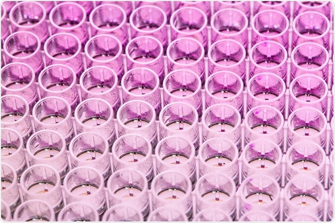
Immunoassays are used in research and hospital laboratories, primarily for monitoring the concentration of specific molecules by utilizing the unique interaction between an antibody and its specific antigen.
 Val3ri0 | Shutterstock
Following the first recorded immunoassays in the 1950s by Yalow and Berson, the industry has grown dramatically, reaching a net worth of $17 billion in 2012.
These methods improve medical intervention, assessing the progression and severity of the illness, and ultimately informing enhanced therapeutic selection. They can also be used in research, identifying the concentration of various biological molecules. Furthermore, in industrial settings, these methods can be applied to monitor contamination and quality of products. There are various types of immunoassay, as described below.
Radioimmunoassay
This traditional immunoassay method utilizes an antigen attached to a radioisotope, bound to an antibody. When the sample is added, it competes with this radioactively-labeled antigen, replacing it and binding to the antibody.
As the concentration of the sample increases, the radioactive antigen is diluted out. Once the unbound antigens are washed away, the amount of radioactivity can be quantified, which inversely indicates the concentration of the target antigen within the sample.
This method utilizes radioactive substances which have various associated health risks, and, therefore, several novel methods have been developed which are much less hazardous.
Enzyme immunoassays
The enzyme-linked immunoassay also referred to as an ELISA, utilizes an enzyme linked to an antibody. Following incubation of this antibody with the target antigen, a substrate is added to the solution which is converted into a colored product by the enzyme. The level of color change can then be quantified to indicate the concentration of antigen.
Fluoroimmunoassay
In this assay, the antibodies are labeled with a fluorescent molecule. Therefore, once they have bound to the target antigen, the fluorescence level can be quantified which indicates the concentration of antigen.
Chemiluminescent immunoassay
Chemiluminescent immunoassays are similar to fluoroimmunoassays and ELISAs, but instead, utilize luminescence to indicate antigen concentration. This method requires an emitter-labeled antibody, which attaches to target antigens.
When this emitter label is excited, it releases light as electrons move to a higher energy state and then fall back down again. This excitation is carried out by a coreactant or catalyst. Overall, the amount of light emitted indicates the amount of antigen present within the sample.
Counting immunoassay
Finally, in the counting immunoassay, beads made of polystyrene are covered with lots of complementary antibodies. The target antigen then binds to these beads, forming clumps. The unbound beads are quantified with a cell counter, which inversely indicates the amount of antigen within the sample.
Further classifications
Within these different types of immunoassay, there are also several different formats. For example, in a competitive immunoassay, the target antigen displaces a labeled antigen from the binding site of the antibody; therefore, the amount displaced indicates the amount of target antigen within a sample.
However, in a non-competitive immunoassay, the antibodies are labeled, and the amount of binding indicates the amount of target antigen within a sample. Such non-competitive assays also exhibit additional advantages such as wider working range and higher precision.
Sources
- https://immunochemistry.com/2016/07/07/back-basics-immunoassay/
- www.lornelabs.com/…/what-is-chemiluminescent-immunoassay
- https://www.sepmag.eu/different-types-of-immunoassays
- https://www.sciencedirect.com/science/article/pii/S0065242301360274
Further Reading
- All Immunoassays Content
- What are Lateral Flow Assays?
- How do Lateral Flow Immunoassays Work?
- Tyramide Signal Amplification (TSA) Methodology
- Tyramide Signal Amplification (TSA) Applications
Last Updated: Jan 18, 2019

Written by
Hannah Simmons
Hannah is a medical and life sciences writer with a Master of Science (M.Sc.) degree from Lancaster University, UK. Before becoming a writer, Hannah's research focussed on the discovery of biomarkers for Alzheimer's and Parkinson's disease. She also worked to further elucidate the biological pathways involved in these diseases. Outside of her work, Hannah enjoys swimming, taking her dog for a walk and travelling the world.
Source: Read Full Article
