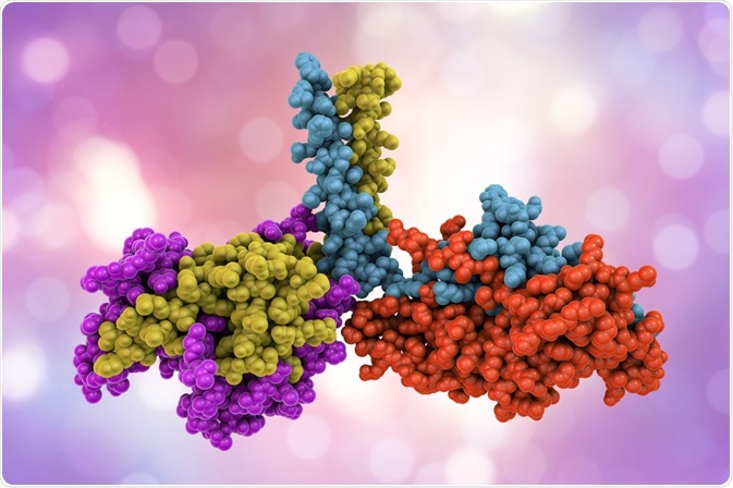
Host cell proteins (HCP) are recombinant proteins that are expressed by a cell of a different species, known as the 'host'. While the presence of HCPs may only be in the part-per-million (ppm) concentration levels, they are highly immunogenic and can have lethal effects on patients if they are not removed. Liquid-chromatography mass spectrometry (LC-MS) is a potential method for the detection of HCPs in biopharmaceutical products.
Skip to:
- Advantages of LC-MS for HCP detection
- Methodology
- Modes of mass chromatography
- Alternative methods for the detection of HCPs
 Kateryna Kon | Shutterstock
Advantages of LC-MS for HCP detection
LC-MS combines the capabilities of two methods: liquid chromatography and mass spectrometry. Liquid chromatography is used to separate different mixtures, while mass spectrometry subsequently identifies the separated molecules. This has several advantages. Firstly, it enables scientists to identify all proteins in a sample, including those that are non-immunoreactive. Secondly, it does not require protein-specific antibodies.
LC-MS has a high sensitivity, is able to detect impurities within the parts per million range, and can provide information both qualitatively and quantitatively. This method can be performed with either one dimensional or two-dimensional LC-MS.
Methodology
Firstly, all the peptides within a sample must be extracted, purified, and fractionated. The sample is then subjected to one dimensional or two-dimensional liquid chromatography followed by mass spectroscopic analysis. The results are entered into a database which contains results for thousands of potential host cell proteins.
Low concentrations
When the host cell proteins are present at very low levels, they may co-elute or precipitate with intense antibody peptides. In such cases, a broader dynamic range may be used, with a different separation phase. Various exclusion and inclusion lists can further enhance the sensitivity of LC-MS.
Modes of mass chromatography
There are several modes that can be used for the detection of mass spectrometry:
- In the shotgun method, the detection and fragmentation of peptide ions are based on the signal intensity.
- In the ‘targeted' method, a set of predetermined fragment ions are detected from the precursor ions that are then anticipated in the survey scan.
- "Directed" methods refer to the selection and fragmentation of peptide ions that are predetermined in the survey scan.
One study showed that a combination method involving directed mass spectroscopy along with two-dimensional fractionation methods was able to identify the highest number of HCPs.
Alternative methods for the detection of HCPs
The methods currently used to detect HCPs include ELISA (Enzyme-linked immunosorbent assay) and western blotting. However, these methods require prior information about HCPs, which is not requi for LC-MS. Also, these methods are time intensive and expensive, and in many cases, they need to be adapted based on the cell type and purification method. In addition, ELISA does not detect HCPs that are non-immunoreactive.
Another method is two-dimensional gel electrophoresis. This method can be coupled to fluorescence microscopy and used in a semi-quantitative manner. However, this method has a limited range and requires the presence of other methods to identify the HCP.
Sources
- Doneanu C.E. & Chen W. (2014) Analysis of Host-Cell Proteins in Biotherapeutic Proteins by LC/MS Approaches. In: Labrou N. (eds) Protein Downstream Processing. Methods in Molecular Biology (Methods and Protocols). doi.org/10.1007/978-1-62703-977-2_25
- JSR Life Sciences. (2017) Comparison of Purification Performance of Amsphere A3 To Other Commercially Available Protein A Resins. biopharma-asia.com/featured-article/comparison-purification-performance-amsphere-a3-commercially-available-protein-resins/
Further Reading
- All Biotherapeutics Content
- How are Host Cell Proteins Removed from Biopharmaceuticals?
- Tracing the Protease Inhibitor AEBSF with RPLC-UV
- Biosimilar Drugs: Overview and Industry Challenges
Last Updated: May 2, 2019

Written by
Dr. Surat P
Dr. Surat graduated with a Ph.D. in Cell Biology and Mechanobiology from the Tata Institute of Fundamental Research (Mumbai, India) in 2016. Prior to her Ph.D., Surat studied for a Bachelor of Science (B.Sc.) degree in Zoology, during which she was the recipient of anIndian Academy of SciencesSummer Fellowship to study the proteins involved in AIDs. She produces feature articles on a wide range of topics, such as medical ethics, data manipulation, pseudoscience and superstition, education, and human evolution. She is passionate about science communication and writes articles covering all areas of the life sciences.
Source: Read Full Article
