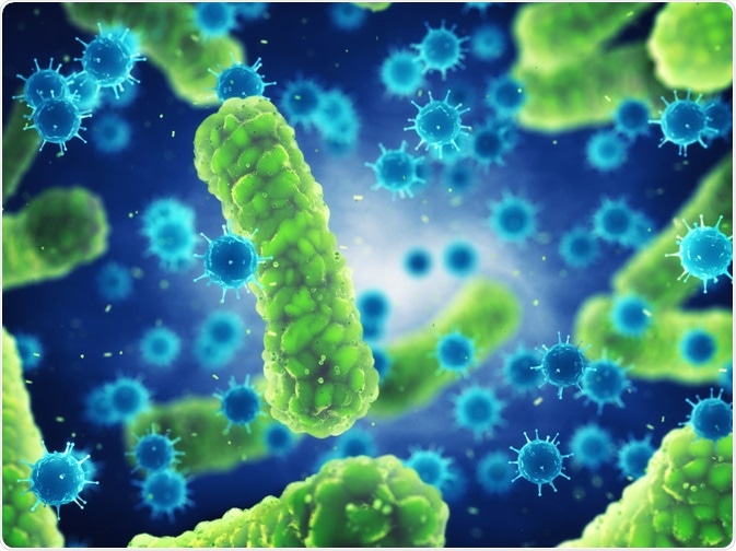
Bacterial infections are an increasing public health threat globally. The ability to rapidly detect infections in potentially life-threatening conditions such as sepsis is vital.
Researchers have presented novel detection techniques that can successfully identify and distinguish healthy and non-viable bacteria within minutes, potentially saving lives and money.

Image Credit: nobeastsofierce/Shutterstock.com
The Important of Bacterial Testing
The modern medical practice relies heavily on bacterial testing. In progressive infections such as sepsis, the mortality rate increases by 8% per hour of delayed treatment.
While up to 30% of those presenting with urinary tract infections are not diagnosed using dipsticks, particularly in those with lower levels of infection.
In either instance, delays in diagnosis can lead to life-threatening consequences as the infection continues to become established. Similarly, delays in identifying cases of contamination in industrial samples can result in adverse economic outcomes.
Typical techniques of bacterial detection include enzyme-linked immunosorbent assay, plate culturing, and polymerase chain reaction. While the tests are credited for their sensitivity and accuracy, practitioners have to wait several days for the results.
Emerging research aimed to address these issues combining mathematical modeling, biology, and engineering principles to create a novel method to detect viable bacteria.
Ultra-fast Technology to Detect Bacteria
The investigation of a cell's electrical characteristic in response to being exposed to external electric fields, known as bioelectricity, is central to the current study.
As a topic, the investigation of bacterial bioelectricity is quite contemporary, but as a result, researchers have discovered that bacteria need a constant resting potential to replicate and use approximately half their energy to maintain this equilibrium.
Previously, fluorophore-mediated and time-lapse microscopy single-cell membrane studies have been used to identify proliferative capacity. One issue with such studies is that membrane potential changes can occur in several situations, creating generalized results if thorough calibrations are not conducted first.
The team of researchers from the University of Warwick, observed the changes in membrane potential and cell proliferation in response to electrical stimulation using a specially-developed device in two species of bacteria, Bacillus subtilis (B. subtilis) and Escherichia coli (E. coli).
A technique called phase-contrast time-lapse microscopy was used to observe when proliferative bacteria absorbed dyed fluorescent molecules used as membrane voltage indictors.
Following the administration of a 2.5-second electrical pulse, an intense fluorescence was observed indicating that the inside of the bacteria cell was more negatively charged compared to the outside.
A proportion of the cells were exposed to ultraviolet light – a known inhibitor of bacterial growth. This impairment of growth was validated using phase-contrast time-lapse microscopy.
Control cells were also identified, and subject to the identical stimulation, irradiated cells became depolarized and the other cells hyperpolarized. This created a distinction between healthy and non-viable bacteria.
This shift was argued to be caused by an alteration in resting membrane potential in damaged cells and anticipated using the extended neuron model in the research.
The researchers treated a mixed culture of the two species of bacteria with an antibiotic, vancomycin, that inhibits B. subtilis proliferation only. Stimulation of both species resulted in hyperpolarization and depolarization of E. coil and B. subtilis, respectively.
The same was observed when the specimens were treated with protonophore or ethanol, resulting in cell damage. This method can be employed in addition to selective culture to identify antibiotic resistance.
Research Implications
James Stratford, the research's lead author, noted that "The system we have created can produce results which are similar to the plate counts used in medical and industrial testing but about 20x faster.
This could save many people's lives and also benefit the economy by detecting contamination in manufacturing processes."
As a result of the research, industrial devices are hoped to be available for clinical and commercial use to promptly identify live bacteria and investigate the effects on antibiotics on cultures.
Rapid Colorimetric Detection
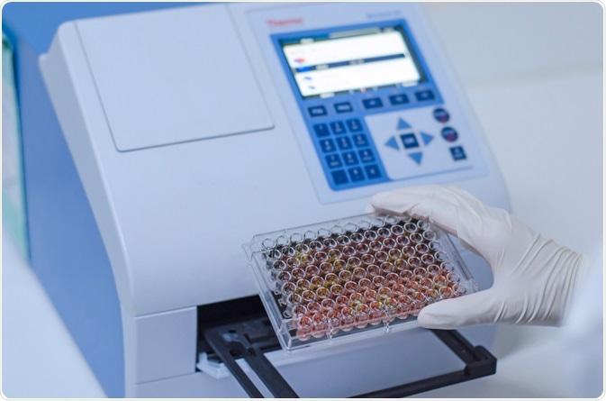
Contemporary research within the field has employed detection techniques involving the use of chemical sensory, including colorimetric-derived detection methods. Such methods are credited for their ease of use and ability to identify the bacteria without the need for additional equipment.

Image Credit: Kallayanee Naloka/Shutterstock.com
A recent study reported on a novel technique called Bacterial Inhibition of GOX-catalyzed Reaction (BIGR) which can rapidly detect a broad spectrum of live bacterial species.
The researchers from Shanghai Jiao Tong University used the technique to identify Salmonella pullorum, Escherichia coli, Enterococcus faecalis, Staphylococcus aureus, and Streptococcus mutants.
The assay utilized each bacteria's metabolism of glucose and enabled the detection of the species using the naked eye. Compared to traditional techniques, the authors noted that the test took 20 minutes to carry out and required only three microliters of sample.
The authors conclude that "the presented platform has great potential for rapid detection of bacteria in clinic and evaluation of bacteria viability. Future development of our study will focus on improving the sensitivity by utilizing a new colorimetric substrate."
Researchers at Gachon University have developed a similar colorimetric technique using nanoparticles. The technique was able to quantify both Escherichia coli and Staphylococcus aureus by monitoring a color change reduction using the naked eye and spectrophotometry within approximately 10 minutes.
The method relies on the activity of chitosan-coated magnetic nanoparticles, which when incubated with a bacteria-containing sample, results in a decrease in peroxidase-like activity.
Like the previous colorimetric technique, the assay presented is suggested by the authors to have great potential for rapidly diagnosing broad-spectrum bacterial infections in house, drastically reducing waiting times and saving lives.
References and Further Reading
- Stratford, J. P., Edwards, C. L., Ghanshyam, M. J., Malyshev, D., Delise, M. A., Hayashi, Y., & Asally, M. (2019). Electrically induced bacterial membrane-potential dynamics correspond to cellular proliferation capacity. Proceedings of the National Academy of Sciences, 116(19), 9552-9557. https://doi.org/10.1073/pnas.1901788116
- Alamer, S., Eissa, S., Chinnappan, R., Herron, P., & Zourob, M. (2018). Rapid colorimetric lactoferrin-based sandwich immunoassay on cotton swabs for the detection of foodborne pathogenic bacteria. Talanta, 185, 275-280. https://doi.org/10.1016/j.talanta.2018.03.072
- Sun, J., Huang, J., Li, Y., Lv, J., & Ding, X. (2019). A simple and rapid colorimetric bacteria detection method based on bacterial inhibition of glucose oxidase-catalyzed reaction. Talanta, 197, 304-309. Doi: https://doi.org/10.1016/j.talanta.2019.01.039
- Le, T. N., Tran, T. D., & Kim, M. I. (2020). A Convenient Colorimetric Bacteria Detection Method Utilizing Chitosan-Coated Magnetic Nanoparticles. Nanomaterials, 10(1), 92. Doi: 10.3390/nano10010092
Further Reading
- All Bacteria Content
- Does Vinegar Kill Bacteria?
- What are Extensively Drug Resistant (XDR) Bacteria?
Last Updated: Apr 15, 2020
Source: Read Full Article
