
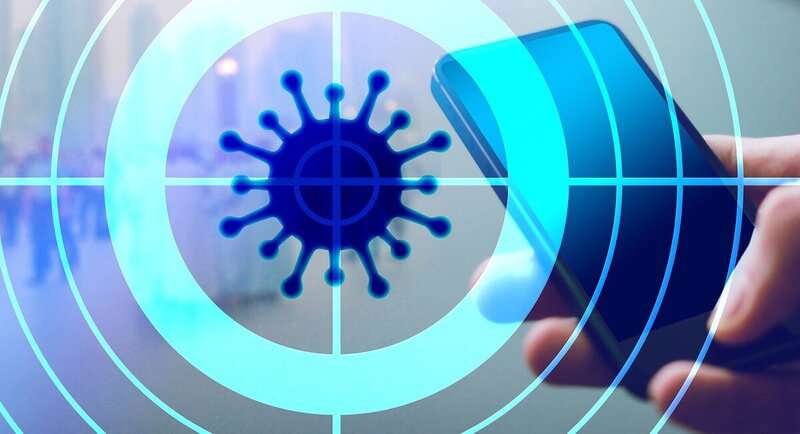
USC researchers are using AI to fuel more confident diagnosis of renal tumors, as well as more customized treatment for cancer patients and patients infected with COVID-19.
Kidney cancer is among the 10 most common cancers. In 2019, the American Cancer Society estimated 73,820 new cases of kidney cancer and 14,770 deaths from this disease. The five-year survival rate reduces from 93% in low-risk groups to 69% in high risk groups of patients with localized kidney cancer. However, following the spread of cancer, these rates plummet to 12%.
For radiologists, a fundamental driver of diagnosing renal cancer remains visual and qualitative, meaning CT scans (images of a mass) are largely evaluated based on individual knowledge and experience. To improve accuracy, this visual analysis has been supplemented by quantitative assessment of renal masses through radiomics, the extraction of quantifiable characteristics from the images.
Researchers at the University of Southern California, including Vinay Duddalwar, director of the USC Radiomics Laboratory and Professor of Clinical Radiology, Urology and Biomedical Engineering at the Keck School of Medicine of USC, and Assad Oberai, Hughes Professor in the Department of Aerospace and Mechanical Engineering and Interim Vice Dean for Research at the USC Viterbi School of Engineering, are combining deep learning with existing contrast CT scanning to help radiologists make more confident diagnoses. Their research was published in the British Journal of Radiology.
The widespread use of contrast enhanced CT, where an intravenous contrast agent like a dye is injected into the tumor and imaged over four distinct points in time, has led to the increased detection of kidney cancers that would have otherwise remained undetected. While many of the tumors identified this way can be labeled benign fairly easily, a significant portion prove more complicated, requiring further invasive testing, the researchers said. Such testing might include biopsies, which might also be inconclusive, pushing many patients to prefer going straight to surgery to remove the tumor in case it is malignant.
“Using a purely visual qualification, 20-25% of all tumors taken out in the U.S. today in the range of 3-5 cm are benign, and didn’t need to come out,” Duddalwar said.
Oberai and Duddalwar recognized this process could be improved by better utilizing existing data. “We wanted to combine what Assad’s group does in deep learning with what my group does in radiomics to improve accuracy of diagnosis,” Duddalwar said.
The researchers also hope such advances could help better understand individual patients’ prognosis in dealing with renal cancer, as well as in addressing diseases such as COVID-19, where individuals report widely varied reactions to infection and treatment.
The research team also includes: from the Keck School of Medicine Associate Professor of Clinical Pathology Manju Aron, Associate Professor of Research Neurology Steven Cen, Executive Director of the USC Institute of Urology Inderbir Gill, medical student researcher at the Department of Radiology Christopher Lau and Assistant Professor in Research Radiology Bino Varghese; and from USC Viterbi computer science student Tomas Angelini and Assistant Professor of Research Radiology and Biomedical Engineering Darryl Hwang.
Contrast Enhanced CT Scans Used to Identify Variations in Tumors
Contrast enhanced CT scans can help diagnose specific cancers, like renal cancer, because of the changes in vascularity seen in such cancers. In a usual workflow, Duddalwar’s group would look at the images of a tumor taken at four different points in time: pre-injection of the contrast agent, 30-40 seconds after injection, 80-90 seconds after injection and then about five minutes after injection. The contrast agent helps identify characteristics related to vascularity, for example, how much blood supply is flowing through the tumor. How early the tumor enhances and washes out compared to the rest of the kidney can help indicate what sort of tumor the patient might have, the researchers said.
“Imagine if you’re sitting at the bank of a river and someone injects a dye further upriver. If the dye gets to where you are quickly, then you know that the current is moving faster. If the dyes spread out, then you know that the flow is turbulent. So you can say a lot about the flow by observing what happens to the dye. Think of the vascular system in the same way. It’s a closed loop fluid system, so if you inject a fluid somewhere, you can watch for it somewhere else,” Oberai said.”For example, if you inject the dye into a blood vessel, but do not observe it downstream, you might be dealing with a tumor that is blocking the vessel and thwarting the flow of blood.”
How the dye diffuses through tissue reveals a lot about the underlying pathophysiology and can help determine a more accurate diagnosis. Instead of recommending more tests and procedures, the deep learning algorithm relies on data collected in the four contrast CT scans. “We are not doing any extra imaging,” Duddalwar said. “We’re using the images already collected and then evaluating them in a different way, so it is no extra expense to the patient or to the healthcare system.” In this way, images collected have the opportunity to convey more data to experts than previously accessible.
Building on Quantitative Evaluations in Radiomics
Radiomics computations can take three to four people about 30-40 minutes to produce results on one patient’s CT scans. An AI algorithm working with the same data can produce results in a matter of seconds.
But efficiency isn’t the most important factor, Oberai said. “More than the time, it’s the effort of experts trying to subjectively figure out where the tumor is, where the boundary is and get the correct margins. What we want to do is save experts’ time for more important tasks, such as evaluating other images and studies, conducting research and teaching and ultimately contributing to improved clinical care through optimized workflow.”
Incorporating deep learning can also help identify new markers that might not otherwise have been discovered. Said Duddalwar: “When you utilize radiomics, you pre-judge, by choosing which element(s) (for example uniformity or asymmetry) you want to evaluate. But with deep learning, you make no such assumption. You let the algorithm figure out what the important characteristic is going to be, which might be an element you never imagined would be significant to diagnosis.”
In the study, the deep learning algorithm demonstrated a 78% accuracy rate in diagnosing the most challenging scans, a rate on par with results produced using radiomics.
Integrating Patient History with Imaging Data
Next the researchers hope to integrate information about a patient’s medical history and clinical examination to help not only improve the accuracy of a diagnosis, but also an individual’s prognosis in treatment.
“We’re looking at using all the imaging information and combining it with clinical data (patient health history, blood tests, symptomology) to make an even more accurate prediction,” Oberai said. “It’s about more than just giving an answer about whether the tumor is benign or malignant, but also producing a number based on all the informational and image inputs that shares how confident the algorithm is about its results.”
He added: “Additionally, we want to be able to have a dynamic model, which can be updated as newer information comes in. For example, a cancer patient might be scanned every three months. We want to see the model updated based on newer data and help better understand the trajectory of the illness for each individual.”
The researchers are looking to apply this beyond diagnosis of renal cancer to its treatment. “We’re trying to find potential markers to help us identify the best treatment straight away instead of wasting months on trial and error,” Duddalwar said. “At the same time, we want to see if deep learning algorithms can help identify which tumors have a better versus worse prognosis for our patients.”
One of the more urgent adaptations the researchers are pursuing is how to leverage this work to better diagnose and treat COVID-19. “Putting together patient symptomology and clinical data with images, you can get a more accurate sense not just of diagnosis but of prognosis. In the case of COVID-19, the data collected can help the model predict how the patient might do—not just whether or not they will recover or get sicker, but also whether or not the patient will need to go to the ICU or require a ventilator.”
The group is going to look at data from COVID-19 patients initially from the USC Health Science campus, which includes the LA County Medical Center. Their research group includes other radiologists, epidemiologists and biostatisticians.
The coronavirus behaves differently in varying locations due to a variety of factors, which are difficult for doctors to access and apply during treatment. However, an algorithm trained on such data can bring in these disparate factors and help link everything together, the researchers said.
Source: Read Full Article