Embryos in pregnancies that end in miscarriage take longer to develop in the womb than those in pregnancies that result in live births, according to new research published today in Human Reproduction.
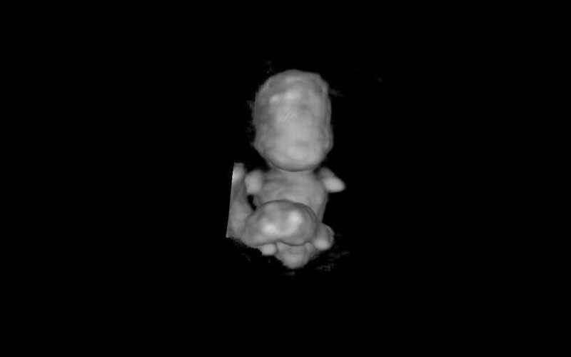
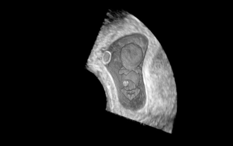
For the first time, researchers in The Netherlands have been able to look at the way embryos develop while pregnancies are ongoing. They used state-of-the-art imaging technology, including 3D ultrasound with high resolution transvaginal probes and virtual reality techniques, to create 3D holograms of the embryo.
This enabled them to assess the overall development of the embryo, including arms and legs, the shape and length of the brain and the curvature of the embryo. 3D ultrasound and virtual reality techniques also enabled them to measure embryonic volume and the distance between the crown of the head and the bottom of the embryo’s buttocks (crown-rump length).
Dr. Melek Rousian, a gynecologist at Erasmus MC, University Medical Center, Rotterdam, The Netherlands, who led the study, said, “We found that in the first ten weeks of the pregnancy, embryos in pregnancies that end in a miscarriage took four days longer to develop than babies that did not miscarry. We also found that the longer it takes for an embryo to develop, the more likely it is to miscarry.
“In the future, the ability to assess the shape and development of embryos could be used to estimate the likelihood of a pregnancy continuing to the delivery of a healthy baby. This would enable health professionals to provide counseling to women and their partners about the prospective outcome of the pregnancy and the timely identification of a miscarriage. This would be particularly useful for couples who have had previous pregnancies that have ended in miscarriage; we might be able to indicate the risk of another miscarriage or maybe offer some early reassurance.”

The researchers collected data from women taking part in the ongoing Rotterdam Periconception Cohort (PREDICT study), a large prospective study embedded in patient care in the department of obstetrics and gynecology at Erasmus MC, University Medical Center, Rotterdam. A total of 611 ongoing pregnancies and 33 pregnancies ending in a miscarriage were included from women recruited to the study between 2010 and 2018 when they were between seven and ten weeks pregnant.
To study the internal and external characteristics and measurements of an embryo, known as embryo morphology, the ability to see the embryo in 3D is important. The researchers used virtual reality to create holograms to look at the embryos’ development and they compared the morphology against established stages of embryo development, known as the Carnegie Stages.
First author of the study, Dr. Carsten Pietersma, a Ph.D. candidate and ultrasonographer at Erasmus MC, said, “Without the aid of 3D and virtual reality, it is far more difficult to examine the development of the embryo. For instance, the 3D virtual reality technology makes it much easier to see the development of the arms and legs. In the Carnegie staging system, the curvature and position of the arms and legs have an important role. Many historic studies have examined the products of miscarriage, but this is the first time we have been able to look at the developing pregnancy while the pregnancy was still intact.”
The Carnegie stages of embryonic development cover the first ten weeks of gestation and run from 1 to 23. Compared to an ongoing pregnancy, a pregnancy ending in a miscarriage was associated with a lower Carnegie stage and the embryo would reach the final Carnegie stage four days later than an embryo from a pregnancy that resulted in a healthy baby. A delay in Carnegie stage increased the likelihood of a miscarriage by 1.5% per delayed stage.
After the tenth week there is no staging system for embryo development, and so the researchers used fetal growth and birth weight to assess development thereafter. They found that a pregnancy ending in a miscarriage was linked to a shorter crown-rump length and smaller embryonic volume.

“We are able to show a significant association between miscarriage and a delay in the early development of the embryo, even if the miscarriage was after ten weeks of gestation,” said Dr. Pietersma.
The researchers adjusted their analyses to take account of factors that could affect pregnancy outcomes such as whether or not the women had been pregnant previously, age, ethnicity, socio-economic status, alcohol use, smoking and use of folic acid or other vitamin supplements.
A limitation of the study is that it includes a relatively small number of pregnancies that ended in miscarriage from a group of women attending tertiary care hospital for preconception and prenatal care and so they may not be representative of the general population. Results of genetic testing following a miscarriage were not available to the researchers, so they do not know if the embryos that miscarried had abnormal chromosome numbers, which could have contributed to the non-viability of the pregnancies.
Dr. Denny Sakkas is Chief Scientific of Boston IVF (U.S.) and the embryology specialist deputy editor of Human Reproduction. He was not involved with the study. He said, “The emotional burden of a miscarriage is incredibly high for women with established pregnancies. This novel study by Carsten Pietersma and colleagues examines the development of embryos in the womb and finds differences in pregnancies that end in miscarriage compared to those that result in live births. The stages of development are calculated from examination of holograms generated from state-of-the-art 3D ultrasound imaging and virtual reality. Use of this technology could prepare patients for an early adverse pregnancy outcome, possibly allowing them to obtain supportive care in case of an adverse outcome.”
More information:
Carsten Pietersma et al, Embryonic morphological development is delayed in pregnancies ending in a spontaneous miscarriage, Human Reproduction (2023). DOI: 10.1093/humrep/dead032
Journal information:
Human Reproduction
Source: Read Full Article
