
Microscopy is the scientific field dedicated to the analysis and imaging of materials too small to be viewed with the naked eye.
The simplest of all optical microscopes is the brightfield microscope, however, scientific advances has lead to the development of more complex, modern technologies that fire photons at a sample and analyze the light scattering patterns in order to generate an image of the sample.
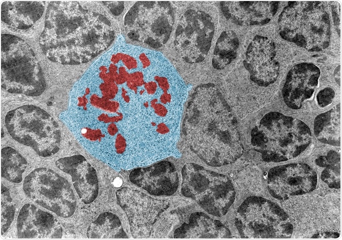 False colour transmission electron microscope (TEM) micrograph of a mitotic cell (blue) surrounded by interphase cells. The chromosomes (red) appear as dark clumps. (Jose Luis Calvo | Shutterstock).
Electron microscopy and X-ray microscopy, which emit electrons or electromagnetic radiation, respectively, are commonly used alternatives to optical microscopes, as electrons possess a much smaller wavelength than photons and thus generate an image of far greater resolution.
X-ray microscopy lies between electron and optical microscopy in terms of resolution, though has the advantage of being usable on living biological samples. Finally, scanning probe microscopy utilizes a physical probe to scan the surface of a sample, detecting minute changes in height on the surface of a sample.
Early microscopes
The first real microscopes were developed in the late 16th century. Several inventors of the microscope have been suggested, including various eyeglasses manufactures in the Netherlands and Italian scientist Galileo Galilei.
In 1665, English scientist Robert Hooke published a book on microscopy called: ‘Micrographia: or Some Physiological Descriptions of Minute Bodies Made by Magnifying Glasses. With Observations and Inquiries Thereupon’.
The book contained a significant number of illustrations of samples observed using a variety of magnifying lenses, including many insects and plants, and even contained the first occurrence of the word ‘cell’.
Aside from biological samples, Hooke also examined the fine edge of a razor and needle points, noting that the seemingly perfectly sharp razor was in fact jagged, while the needle appeared blunt at such a scale.
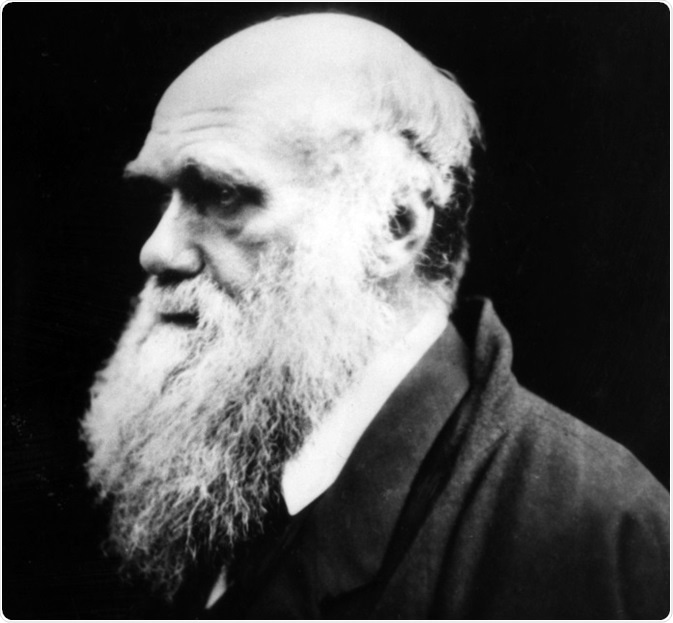 Charles Darwin also made great use of optical microscopes in the late 19th century. He undertook major research work on barnacles, using microscopy to describe their internal structure.
Darwin was considered an expert on microscopy during his lifetime, and even sold a microscope of his own design. Darwin’s microscopy work formed a key part of his theory of speciation, categorizing various extant and extinct species of barnacles, using fossils, into groups. Darwin went on to compare these barnacles with crustaceans, providing compelling evidence regarding the theory of evolution by natural selection.
Modern microscopes
The first scanning electron microscope was created for commercial sale by English scientists Professor Sir Charles Oatley and Dr. Gary Stewart in 1965, working at Cambridge University. They built the original instrument from scratch in the mid 1950’s to study the mechanisms of ion sputtering.
Over the next decade various improvements were made by Oatley’s team, including the incorporation of magnetic lenses to better aim and focus the electron beam.
Electron microscopes possess a resolution of as low as 0.1 nm, allowing for the imaging of objects such as virus’ and even strands of DNA. Electrons are produced and emitted from an electron gun, which passes through several lenses and apertures to focus the electron beam before reaching the sample. The whole chamber containing the electron beam and sample are kept at low pressure in order to minimize interference from gas molecules.
 Konstantin Kolosov | Shutterstock
Source
- Charles Darwin and Robert Brown – their microscopes and the microscopic image.
- Between the Beagle and the barnacle: Darwin’s microscopy, 1837–1854.
- Persistence pays off: Sir Charles Oatley and the scanning electron microscope.
- Dr Robert Hooke.
- Optical Microscopy Review Article.
- A critical review of orientation microscopy in SEM and TEM.
- Darwin Correspondence Project.
- Scanning Electron Microscopy.
Further Reading
- All Electron Microscopy Content
- Electron Microscopy: An Overview
- Applications of Electron Microscopy
- Advantages and Disadvantages of Electron Microscopy
- What is Transmission Electron Microscopy?
Last Updated: Feb 28, 2020

Written by
Michael Greenwood
Michael graduated from Manchester Metropolitan University with a B.Sc. in Chemistry in 2014, where he majored in organic, inorganic, physical and analytical chemistry. He is currently completing a Ph.D. on the design and production of gold nanoparticles able to act as multimodal anticancer agents, being both drug delivery platforms and radiation dose enhancers.
Source: Read Full Article