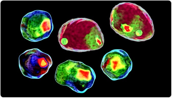
A team headed by Hideharu Mikami from the University of Tokyo recently determined an imaging method based on optomechanics that overcomes the performance and utility of imaging flow cytometry (IFC).

Image Credit: Kateryna Kon/Shutterstock.com
The method enables high throughput imaging of single cells traveling at >10,000 cells sec-1 without compromising sensitivity or resolution and enabling robust statistical analysis and accurate classification of cells.
Traditional IFC offers several advantages over traditional flow cytometry by providing quantitative image data. This data permits the characterization of single cells across heterogeneous populations. The big data resulting from IFC methodology is relevant in a time where deep learning necessary to improve clinical decision-making in the biomedical and clinical settings.
The limitations of traditional imaging flow cytometry
IFC is particularly effective for the enumeration of a range of macromolecules including proteins, nucleic acids, the analysis of cell-cell interaction, and the characterization of DNA damage and repair. However, the performance and utility of IFC suffer as a result of attempts to increase throughput, spatial resolution, and sensitivity.
By increasing the flow speed for high-throughput, a shortened integration period results, enabling the collection of blur-free images; however, the effect is reduced sensitivity or pixel resolution to combat this.
To overcome this, time delay and integration (TDI) with an image sensor based on a charged coupled device (CCD) has been used. TDI accumulates several exposures of cells in flow, with several rows of CCD photosensitive elements. However, this limits throughput as the CCD readout rate is decreased.
An additional problem suffered by CCD is readout noise, which limits its sensitivity of detection. Some attempts to overcome the trade-off are single-pixel imaging and use of a complementary metal-oxide semiconductor (CMOS) image sensor – but these, in turn, limit sensitivity.
The unifying trait of these compensatory techniques is the compromise of one of the key parameters in favor of others. This prevents applying IFC to niche applications, which motivates the study by the team at the University of Tokyo.
A novel optomechanical imaging method
Mikami et al. refer to their improved imaging method as virtual freezing fluorescence imaging (VIFFI). Crucially, tradeoffs are circumvented whilst high throughput (>10,000 cells sec-1), spatial resolution at approximately 700nm, and high sensitivity are ensured.
The method is based on freezing a flowing cell on the image sensor by canceling the motion of a single cell to increase the exposure time of the image sensor. This produces a fluorescence image with a high signal-to-noise ratio (SNR).
The essential elements of VIFFI flow cytometry is an excitation beam scanner that scans over the field of view (FOV) and simultaneous timing of the image senses exposure and the excitation beams illumination. In combination, virtual freezing translates to 1000 times greater times for signal integration on the image sensor. This results in microscopy-level fluorescence imaging of cells at 1 m s-1.
The methodology behind the group’s technique involved the optical design of the VIFI flow cytometer, the performance of an excitation beam scan to extend the exposure time for the fast-flowing cells, acquisition of data, and the processing of the digital images and deep learning.
Applying virtual freezing fluorescence imaging
The group used two colors. The team pointed out that several colors can be used for fluorescence imaging when dichroic beamsplitters are used. The group also remark on the compatibility of VIFFI flow cytometry with advanced image-sensor-based fluorescence microscopy technique – enabling super-resolution fluorescence.
Additionally, the technique enables the flow speed to be varied so long as it conforms to the tradeoff relationship between the FOV, exposure time, and flow speed. Along with this advancement, machine learning will allow the depth of data analysis achieved to be expanded. Similarly, as advances in image sensor technology are achieved, high-throughput and number of fluorescence color options are hoped to be increased. VIFFI also enables the depth of field to be expanded. This is useful for FSIH imaging and imaging of large cells.
Owing to sensitivity spatial resolution on high throughput of this fluorescence imaging, the applications in biology, pharmacy, and medicine are expanded. Firstly, high throughput FISH analysis is capable; this technique is especially useful in diagnosis, identification, and detection of minimal residual disease. Traditional microscopy is insufficient as screening of large populations of cells is not possible.
As proof of concept, the team demonstrated large scale analysis based on morphological phenotype. This suggests that this technique is invaluable when determining genotype-phenotype relation. Specifically, the high spatial resolution enables accurate characterization of key features such as area and perimeter of each cell and intracellular organelles.
The group also illustrated through imaging of mutants of C. reinhardtii, VIFF I flow cytometry can be used for evaluating mutagenesis.
Finally, VIFFI flow cytometry can both detect and count circulating tumor cells (CTCs) from heterogeneous blood samples. Single CTC’s and clusters can be visualized and counted; this is expected to aid the study of the correlation between cluster size and metastatic potential/spread of CTCS.
Source
- Mikami, H. et al (2020) Virtual-freezing fluorescence imaging flow cytometry. Nature Communications. DOI: 10.1038/s41467-020-14929-2.
Further Reading
- All Flow Cytometry Content
- Flow Cytometry Methodology, Uses, and Data Analysis
- Flow Cytometry Techniques used in Medicine and Research
- Flow Cytometry History
- Fluorescence-Activated Cell Sorting
Last Updated: Aug 5, 2020

Written by
Hidaya Aliouche
Hidaya is a science communications enthusiast who has recently graduated and is embarking on a career in the science and medical copywriting. She has a B.Sc. in Biochemistry from The University of Manchester. She is passionate about writing and is particularly interested in microbiology, immunology, and biochemistry.
Source: Read Full Article
