
Biological systems are incredibly complex, with numerous types of cells working together in tissues and organs, performing processes necessary to sustain life. To fully understand these processes and their implications a variety of techniques have been developed and refined over the years.
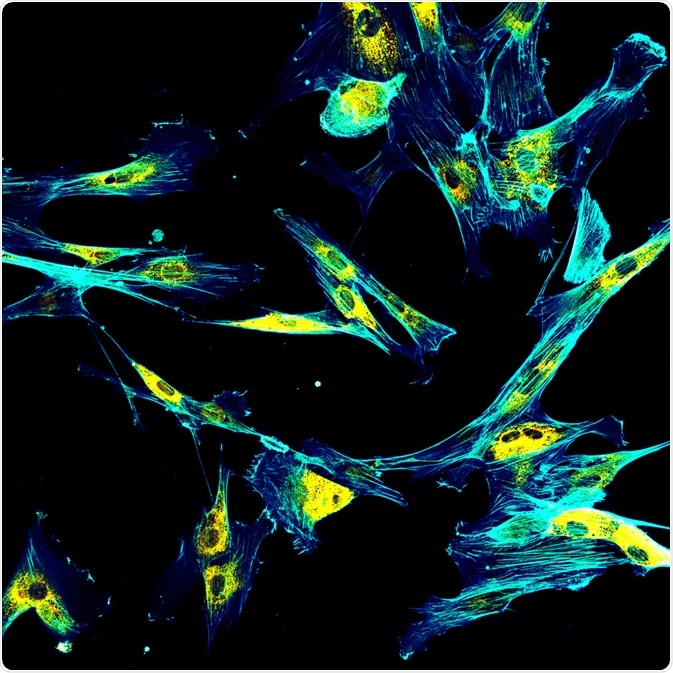
Image Credit: DrimaFilm/Shutterstock.com
Immunofluorescence microscopy is one such technique that is widely employed by researchers. It is an incredibly reliable technique, which uses specific antibodies to target proteins linked to fluorophores (fluorescent chemical compounds) to produce highly detailed images of cell structures and activity.
Issues with conventional IF microscopy
However, immunofluorescence (IF) microscopy is not without its limitations. Due to the spectral overlap of the fluorophores, conventional IF methods have finite multiplexing capabilities. Two points that are <200 nm apart are not distinguishable due to wavelength interference (that is, their fluorescence halos overlap).
To visualize structures at this scale fluorescence intensity can be intentionally depleted, creating narrower fluorescent points (as in STED microscopy) which creates a crisper image, or an image can be reconstructed from several fluorescence points in molecules that are far apart, which are individually activated then switched off so that their halos don’t overlap (PALM and STORM microscopy.)
IF microscopy is generally limited to being able to visualize only four fluorophores at any one time, meaning insufficient information is produced about the range of structures and cellular activity that exist in complex and dynamic biological systems.
One method that has been developed in recent years to overcome this problem is PAINT, which stands for point accumulation for imaging in nanoscale topography.
What is PAINT?
PAINT is a process that uses fast and transient dyes to capture several fluorescence points at once. It was originally developed as a dye-based alternative strategy for super-resolution imaging. Instead of the stochastic photo-activation of a permanently bound fluorophore, PAINT relies on the stochastic binding of a fluorescent ligand.
The technique was first developed by Sharonov and Hochstraser in 2006 using Nile Red, a lipophilic dye, and using the dye to target large unilamellar vesicles (LUVs.) They were able to assemble a super-resolution image which was enabled by intermittent collisions of the dye with the LUVs, localization, and subsequent photobleaching. It was Sharonov and Hochstraser who coined the term ‘point accumulation for imaging in nanoscale topography’.
In this iteration, it was the nature of the dye’s hydrophobic interaction with the membrane bilayer that produced the desired results, rather than the specific actions between the imager and target. As this could not be reversed, an additional step where the sample was bleached was necessary. This was not dynamic enough, but the concept had been proven.
From the initial concept, PAINT was further developed, which allowed to dynamic imaging of other target biomolecules continuously and stochastically with fluorescent ligands in solution, and eventually, custom fluorophores that transiently bind to molecules were developed.
The “blinking” image produced using PAINT allows the center of the fluorescence peak to be captured and resolved with a higher degree of accuracy than by conventional dye-based strategies. This means that an otherwise blurry image becomes much sharper, overcoming a common problem with conventional microscopy.
Whereas conventional immunofluorescent techniques involve sequentially staining target biological structures and repeatedly imaging them to produce a composite image, PAINT is a much more efficient process. It removes the need to repeatedly incubate overnight with different antibodies, subjecting the sample to washing with harsh chemicals, which is laborious and time-consuming. Point accumulation for imaging in nanoscale topography represents a major step forward in the imaging capabilities of super-resolution microscopy in real-time.
Further Developments in PAINT
Recently, research teams have been developing methods that utilize the principles of PAINT. The technique has been combined with specifically marked DNA oligonucleotides to produce DNA-PAINT. DNA-PAINT was combined with PAINT principles in an exchangeable manner to produce Exchange-PAINT by Dr. Yin et al. in 2014. qPAINT and FRET-PAINT are also recent developments. The principle of the method is being expanded upon in many novel ways.
One problem does exist in PAINT however: the current library of available antibodies is limited. To fully exploit the potential of this technique, new and novel antibodies (for example, those which could be produced by the advent of semi-synthetic organisms) will have to be produced. However, due to the dynamic nature of the method, this represents a relatively minor stumbling block that is being remedied by advances in other fields.
PAINT represents a revolutionary method for imaging and studying the complex, dynamic processes that exist within biological systems.
Sources
Nieves, D.J et al. (2018) DNA-Based Super-Resolution Microscopy: DNA-PAINT, Genes (Basel) Vol. 9 Issue 12 pg. 621.
https://doi.org/10.3390/genes9120621
Sharonov A., Hochstrasser R.M. (2006) Wide-field subdiffraction imaging by accumulated binding of diffusing probes. Proc. Natl. Acad. Sci. The USA. Vol. 103 Issue 50 pgs. 18911–18916.
https://doi.org/10.1073/pnas.0609643104
Wang, Y et al. (2017) Rapid Sequential in Situ Multiplexing with DNA Exchange Imaging in Neuronal Cells and Tissues Nano Lett. Vol. 17 Issue 10 Pgs. 6131-6139
https://doi.org/10.1021/acs.nanolett.7b02716
Last Updated: Jan 17, 2020

Written by
Reginald Davey
Reg Davey is a freelance copywriter and editor based in Nottingham in the United Kingdom. Writing for News Medical represents the coming together of various interests and fields he has been interested and involved in over the years, including Microbiology, Biomedical Sciences, and Environmental Science.
Source: Read Full Article