The tissue-specific immunity of the lungs
The immune cells of the lung
Case study: Bacterial pneumonia
Conclusion
References
Further reading
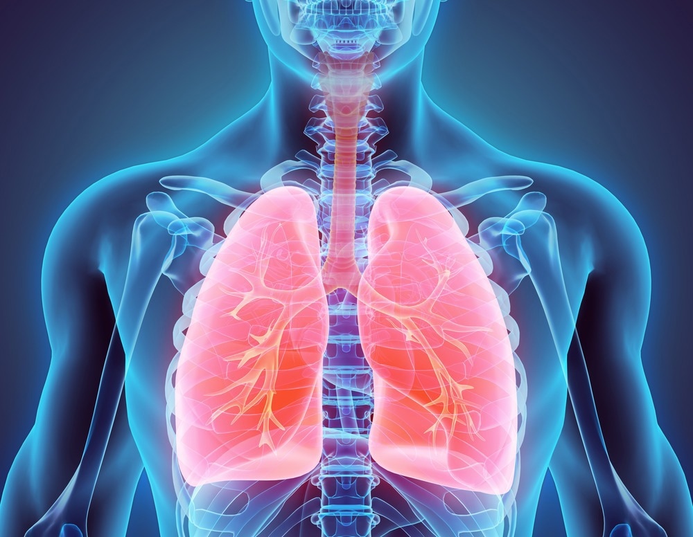
The lungs play a vital role in respiration but also as an immune organ. The tissue-resident immune cells in the lungs act as the first line of defense against pulmonary infections. Although tissue-resident memory T cells are key components of lung immunity, tissue-resident innate immune cells also play a significant role in pulmonary diseases such as influenza, bacterial pneumonia, asthma, and inflammatory disorders.

Image Credit: MDGRPHCS/Shutterstock.com
The lungs are specialized organs for gas exchange, representing one of the largest epithelial surfaces in direct contact with the external environment. They also serve as immune organs, fostering both innate and adaptive immune cells. Due to their direct contact with the outer environment, the lungs serve as a primary target for a diversity of airborne pathogens, toxic compounds, and allergens causing acute respiratory distress syndrome (ARDS), pneumonia, and acute lung injury (ALI).
The lung's immune system plays a crucial role in protecting the body from all these respiratory pathogens while tolerating small particulate matter and mechanical forces from respiration. Recent studies have shown that a complex network of non-recirculating immune cells residing within lung tissue is responsible for maintaining a balance between immunity and tolerance.
The tissue-specific immunity of the lungs
The pulmonary immune system is composed of different compartments. The upper respiratory tract serves as a mucosal and glandular component dominated by IgA antibodies, while the peripheral airways lack mucosal tissue and are dominated by IgG antibodies.
The peripheral airways are constantly in contact with broncho-alveolar cells (BACs), which are mainly composed of alveolar macrophages (AM) and lymphocytes (also found in a compartment of the respiratory tract epithelium). The bronchus-associated lymphoid tissue (BALT) is another compartment of the respiratory lymphoid cells (RLCs), and it comprehends organized lymphoid tissues present inside the bronchial walls.
BALT disappear after adolescence; adults present it only during chronic inflammatory diseases, where it is called inducible BALT (iBALT). B cells are the major cell population of BALT, but T cells and dendritic cells (DCs) are also present. It also has high endothelial venule (HEV), serving to transport lymphocytes and antigens between the lungs and the circulation.
The other compartment comprises BACs, obtained through broncho-alveolar lavage fluid (BALF) from the peripheral airways contain AMs, innate lymphoid cells (ILCs), and DCs, which protect against inhaled pathogens, toxicants, and allergens. These cells act as antigen-presenting cells (APCs), secreting several cytokines and chemokines to regulate innate and adaptive immunity.
The immune cells of the lung
Alveolar macrophages are considered the prototypic lung-resident immune subset. Following the first breath, macrophages differentiate into long-lived alveolar macrophages (AMs) and interstitial macrophages (IMs). AM are considered anti-inflammatory cells with an important role in phagocytosis of particulate matter, dying cells, and cellular debris, maintaining immune homeostasis through the production of TGF-β and subsequent induction of FoxP3 regulatory T cells (Treg).
During infection, their phagocytic activity is increased. However, AMs still have a role in limiting inflammation by producing TGF-β, prostaglandin-E 2, and platelet-activating factor. Failures in phagocytosis are associated with chronic obstructive pulmonary disease (COPD) and asthma. On the other hand, IMs have been shown to limit Th2 responses through secretion of IL-10. Both AMs and IMs are involved in tissue repair.
Innate lymphoid cells (ILCs) in the lung play a role in immunosurveillance and infection control. In mice infected with H1N1 influenza virus, ILC1s are activated and produce interferon γ (IFN-γ) and tumor necrosis factor α in the first three days of infection. ILC2s constitutively express IL-5 and are induced to secrete IL-13 under inflammatory stimuli, resulting in eotaxin production, thereby controlling eosinophil accumulation.
ILC3s produce Interleukin-17, protecting from some intracellular bacteria, such as M. tuberculosis, as well as extracellular bacteria and fungi. Despite this, ILC3s can also drive inflammatory responses. For example, IL17+ ILC3s are enriched in bronchoalveolar lavage samples in humans with severe asthma. Unlike ILCs, NK cells constantly recirculate and may contribute to chronic inflammatory diseases since they have been associated with COPD and asthma by producing inflammatory cytokines.
Regarding DCs, their heterogeneity in the lung is still being studied. There are plasmacytoid DCs (pDCs) and myeloid DCs, which are subdivided into cDC1 and cDC2. Lung DCs do not appear to self-renew; they arise from precursor cells and persist in the lung for up to 8 weeks. They have been shown to elicit different functions against a variety of stimuli.
Data from experimental mice models challenged with house dust mites (HDM) suggest that these cells play important roles in T-cell response in lung inflammation. In this context, allergens are taken up by CD11b+ cDC2s, causing Th2 and Th17 responses. In contrast, CD103+ cDC1s limit this allergic inflammation produced by chronic HDM exposure via the production of IL-12. CD103+ cDC1s also induce Treg cells in response to HDM via retinoic acid signaling.
Case study: Bacterial pneumonia
Different tissue-resident cells act in pathogenic situations. The best example is AMs in the pathogenesis of bacterial pneumonia. AMs during gram-negative bacterial pneumonia produce TNF-α, which induces granulocyte-macrophage colony-stimulating factor (GM-CSF) in airway epithelial cells (AECs), generating proliferative signaling via autocrine stimulation and restoring epithelial barrier. Although during bacterial pneumonia, infiltrating peripheral macrophages replace the resident macrophages. The AM-mediated elimination of apoptotic cells decreases its phagocytic potential against bacterial pathogens, increasing bacterial growth in the lungs.
In mice, pulmonary NK cells protect against K. pneumoniae-induced pneumonia by secreting IFN-γ and IL-22, which enhances neutrophil infiltration and ALI. However, ILC3s later increase IL-17 levels to improve phagocytosis of the pathogen and the resolution of the Gram-negative bacterial pneumonia-induced ALI.
Conclusion
Ongoing research aims to develop more effective treatments for lung diseases, such as pneumonitis, fibrosis, and lung cancer, by harnessing the role of tissue-resident immune cells. Some of these studies are being carried out to gain insights into their functions and to understand their intricate interactions with the lung microbiome in both health and disease.
References
- Ardain, A., et al. (2019). Tissue‐resident innate immunity in the lung. Immunology, 159(3), pp. 245–256. https://onlinelibrary.wiley.com/doi/10.1111/imm.13143
- Kumar, V. (2020). Pulmonary innate immune response determines the outcome of inflammation during pneumonia and sepsis-associated acute lung injury. Frontiers in Immunology, 11. https://www.frontiersin.org/articles/10.3389/fimmu.2020.01722/full
Further Reading
- All Immunology Content
- What is Immunology?
- What does it mean to be Immunocompromised?
- Classical Immunology
- The Immunological Functions of Red Blood Cells
More…
Last Updated: Jun 16, 2023

Written by
Deliana Infante
I am Deliana, a biologist from the Simón Bolívar University (Venezuela). I have been working in research laboratories since 2016. In 2019, I joined The Immunopathology Laboratory of the Venezuelan Institute for Scientific Research (IVIC) as a research-associated professional, that is, a research assistant.
Source: Read Full Article
