
Skeletal muscle cells facilitate voluntary movements such as walking, running, and other forms of exercise. They are comprised of thin, tubular cells called muscle or myofibres due to their threadlike appearance. A single muscle is comprised of hundreds to thousands of myofibres each of which is comprised of units of contractile machinery, called sarcomeres.
Sarcomeres contract and lengthen and during exercise; eccentric contraction cause fibers to be forcibly lengthened during contraction can result in overstretching. Physical exercise induces mild musculoskeletal injuries that need rapid and efficient repair to preserve muscle homeostasis.
However, subsequent injury produces reduced damaging effects because of adaptation. This adaptation is in consequence of muscle repair, and several proposed mechanisms have since been unearthed since the discovery of damage to the muscle fiber.
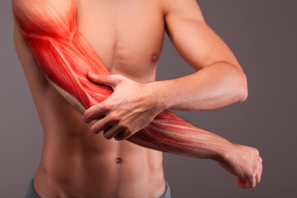
Image Credit: BigBlueStudio/Shutterstock.com
When does muscle regeneration occur?
Muscle regeneration in response to injury typically initiates during the first week after injury, peaking at two weeks, and gradually slowing between three and four weeks.
The role of satellite cells in muscle regeneration
Resident muscle stem cells, also known as satellite cells, are located between the plasmalemma of myofiber on the basal lamina. These specialized cells possess a potential for muscle regeneration following injury. After muscles are injured, satellite cells become rapidly activated from a resting or quiescent state.
They subsequently differentiate to assume myoblasts and proliferate and fuse or with existing myofibers (forming myotubes) to regenerate muscle. A small population of these activated cells then revert to the quiescent state where they can self-renew and ensure the stem cell pool is maintained.
The concentration of satellite cells is elevated in type I muscles (slow-twitch; more efficient at using oxygen to generate more ATP for continuous, extended muscle contractions) and is lower in older people. even after repeated injury, the concentration of satellite cells is maintained at a constant.
The recovery process
- Non-inflammatory degenerative phase: autolysis of the myocytes occurs
- Inflammatory degenerative phase: phagocytosis begins due to the migration of macrophages is. These eliminate damaged fibers. An increase in neutrophil concentration occurs through the increase in proteolysis of the extracellular matrix. This takes place between one and six hours. Collagenase, an important proteolytic enzyme, is secreted by fibroblasts. Other inflammatory cells secrete cytokines and growth factors. Nonsteroidal anti-inflammatory drugs affect the repair process in damaged muscles -reducing the associated inflammation, swelling, and pain that peak 1–2 days post-injury.
- Regenerative phase: this occurs quickly with closed injuries. This process begins with the activation of satellite cells in the basal lamina (if it remains intact)
- Maturation phase: satellite cells differentiate; this is stimulated by insulin-like growth factor (IGF). During this phase, gene expression is altered by the fusion of cells causing muscle-specific protein formation. Myogenesis follows the stages of embryonic development. First, fast-twitch filaments are produced from undifferentiated embryonic myosin filaments. Slow-twitch myosin is subsequently produced, and because of innervation, slow-twitch muscle is produced. Creatinine kinase activity also increases
Regenerative phase
In adult muscle cells, quiescent satellite cells express the box protein 7 (PAX7), on the activated cells upregulate myogenic regulatory factor MYOD and proliferate. When cells undergo differentiation into myocytes, most satellite cells downregulate PAX7 and maintain the expression of MYOD. This allows the cells to initiate the process of myogenesis via upregulating the protein myogenin.
In myoD-knockout mice, there is a reduced regeneration ability following muscle injury as a consequence of impaired population expansion and differentiation; this indicates that MYOD plays an essential role in activating satellite cells during the initial phase of regeneration.
How the activation of satellite cells is regulated has long been considered a fundamental question in muscle biology. Towards the end of the 20th century, researchers determined that muscle extracts are capable of activating and proliferating cultured muscle cells; this subsequently prompted the exploration of factors in these extracts from intact or crushed muscle tissues that contribute to muscle regeneration.
As a result, several growth factors that promote the activation and proliferation of satellite cells were unearthed from muscle tissues. Growth factors modulate satellite cell activity and are secreted from several cells located in the institutional spaces. These include macrophages, mesenchymal progenitors, and fibroblasts.
In a recent study, researchers sought to investigate the regulation of satellite cell activation using isolated individual myofibres in a floating culture model. In doing so, researchers could view myofibre extracts and quiescent satellite cells associated with myofibres in the absence of interfering effects of the interstitial cells.
This revealed that damaged yofiber-derived factors (DMDFs) are capable of stimulating satellite cells and cause them to transition from the G0 (quiescent) to the G1 stage of the cell cycle. The group also identified metabolic enzymes (such as GAPDH) that function as DMDFs to activate satellite cells ex vivo and stimulate the expansion of satellite cells during muscle regeneration in vivo.
Taken together, this group demonstrated that damaged myofibers directly signal to residents’ stem cells following muscle injury to expedite the first step of tissue regeneration. In addition, DMDFs function as biomarkers for muscle damage as well as initial trigger proteins that are involved in the activation of satellite cells.
More recently than this, researchers have discovered a previously unidentified repair mechanism that is triggered after muscle injury. Researchers demonstrated a repair process that is independent of muscle stem cells (cell-autonomous) after using mouse models.
In response to injury, mouse muscle was found to trigger a signaling cascade that results in the attraction of my nuclei to the damaged site as the consequence of microtubule and dynein assembly. These movements were found to accelerate the repair of the sarcomere and cause local delivery of messenger RNA for cellular reconstruction.
Researchers described this as a cell-autonomous protective mechanism, a form of myofibre self-repair, which represents an alternative model for understanding how the morphology of muscle is restored in health and disease.
This finding challenges the skeletal muscle and homeostasis and regeneration dogma which is widely assumed to be mediated by satellite cells. The work opposes this stem cell-centric view, demonstrating that muscle integrity is also maintained through an alternative myofibre autonomous repair mechanism. At the heart of this mechanism is a nuclear migration for local delivery of messenger RNA is required for the production of protein and repair of damaged sarcomeres.
References:
- Roman W, Pinheiro H, Pimentel MR, et al. (2021) Muscle repair after physiological damage relies on nuclear migration for cellular reconstruction. Science. doi: 10.1126/science.abe5620.
- Tsuchiya Y, Kitajima y, Masumoto H, et al. (2020) Damaged Myofiber-Derived Metabolic Enzymes Act as Activators of Muscle Satellite Cells. Stem Cell Reports. doi: 10.1016/j.stemcr.2020.08.002.
- Sambasivan R, Yao R, Kissenpfennig A, et al. (2011) Pax7-expressing satellite cells are indispensable for adult skeletal muscle regeneration. Development. doi: 10.1242/dev.067587.
- Chargé SB, Rudnicki MA. (2004) Cellular and molecular regulation of muscle regeneration. Physiol Rev. doi: 10.1152/physrev.00019.2003.
Further Reading
- All Muscle Content
- How to Reduce Muscle Soreness After Training
Last Updated: Jan 11, 2022

Written by
Hidaya Aliouche
Hidaya is a science communications enthusiast who has recently graduated and is embarking on a career in the science and medical copywriting. She has a B.Sc. in Biochemistry from The University of Manchester. She is passionate about writing and is particularly interested in microbiology, immunology, and biochemistry.
Source: Read Full Article