
Introduction
Techniques to Uncover Brain Maps
Lesion Studies and Their Influence in Neuropsychology
The Utility of Functional Neuroimaging in Brain Mapping
Brain Mapping at a Cellular Level: the Connectome
Where will Brain Maps Lead us in the Future?
References
Further Reading
Neuropsychology is a subdivision of psychology and neurology which refers to the relationship between an individual’s behavior and the physiological processes of the brain and central nervous system.
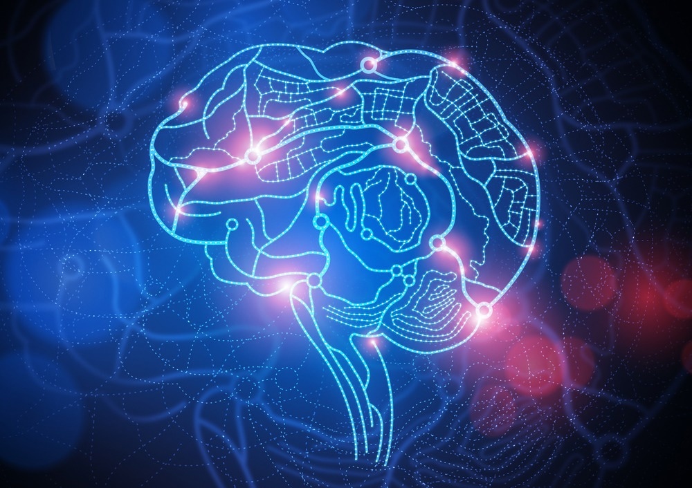
Connections in the brain. Image Credit: solarseven/Shutterstock.com
This has led to an exponential interest in brain mapping, which aims to uncover the relationship between the structure and function of the brain. The ability to pinpoint which structures in the brain are responsible for cognitive function would be an invaluable tool for a range psychiatric disorders and cognitive impairments.
With the advancement in neurosurgery tools, neuroimaging technology and the advent of machine learning, using brain maps to predict behaviors is closer than ever before.
Techniques to uncover brain maps
Various tools developed over the last century have alluded to localization of function in the brain. This marriage of cognitive psychology and functional brain imaging has included neuroimaging techniques such as: functional magnetic resonance imaging (fMRI), positron emission tomography, electroencephalography, electrocorticography and near-infrared spectroscopy, a recently emerging tool.
Electrophysiologic techniques including deep brain stimulation may also be used to probe neuronal function in distinct brain regions. Used in isolation or a multi-modal approach, these tools are used to attempt to decipher neuronal structures, cells and regions which are responsible for human behavior.
Lesion studies and their influence in neuropsychology
Original brain mapping studies have investigated the impact of traumatic brain injury and brain lesions in distinct regions and their impact on cognitive function. Such studies were pivotal in the field of neuropsychology and provided the first insights into the neural basis of behavior.
Seminal articles in the mid to late 20th century included influential works that revealed hippocampal lesions were associated with impaired memory and injuries to the Wernicke area were responsible for language comprehension deficits. Although such studies have continued to reveal evidence for the localization of learning, cognitive control, social behavior and memory in the brain, they are limited by their size, and variability between patient lesions is difficult to control since they cannot be experimentally produced.
The utility of functional neuroimaging in brain mapping
The emergence of fMRI scanning has been invaluable in the quest to develop neural atlases of behavior. These methods examine the association between cognitive processes and activity in neurons, brain regions, or networks.
Principally, fMRI measures changes in blood flow which translate to cellular activity and theoretically provide a functional brain map. Tasked-based fMRI enhances this map by having subjects perform tests during an MRI scan to identify stimulus-evoked patterns of cellular activity. Prominent examples include the performance of working memory tasks which led to activation in the prefrontal cortex of healthy subjects, reinforcing this structure as fundamental in memory.
Adversely, however, prefrontal cortex activation declines during aging, and was found to be significantly reduced in frontotemporal dementia patients. Other examples include studies that have exposed subjects to a range of facial expressions during scanning, which identified the importance of the amygdala in social behavior and recognition of facial expressions and emotion.
In autism, which features impaired social interaction and emotion processing, trials have highlighted altered connectivity and activation of the amygdala. This evidence reinforces the utility of functional neuroimaging studies in mapping behavior not only in healthy individuals but also by uncovering altered function in brain regions involved in cognitive and psychological disorders.
Whether this provides sufficient evidence to utilize fMRI findings as biomarkers remains controversial. FMRI results should be interpreted with caution since they provide measures of a surrogate of brain activity via blood flow and changes in blood oxygen. Some have even argued against the notion that the brain has a fixed neural architecture. Moreover, in contrast to lesion studies, these techniques cannot differentiate between regions that are involved in a function from those which are necessary. The combination of fMRI use in lesion studies may overcome this issue to provide a more thorough localization of behaviors in the brain.
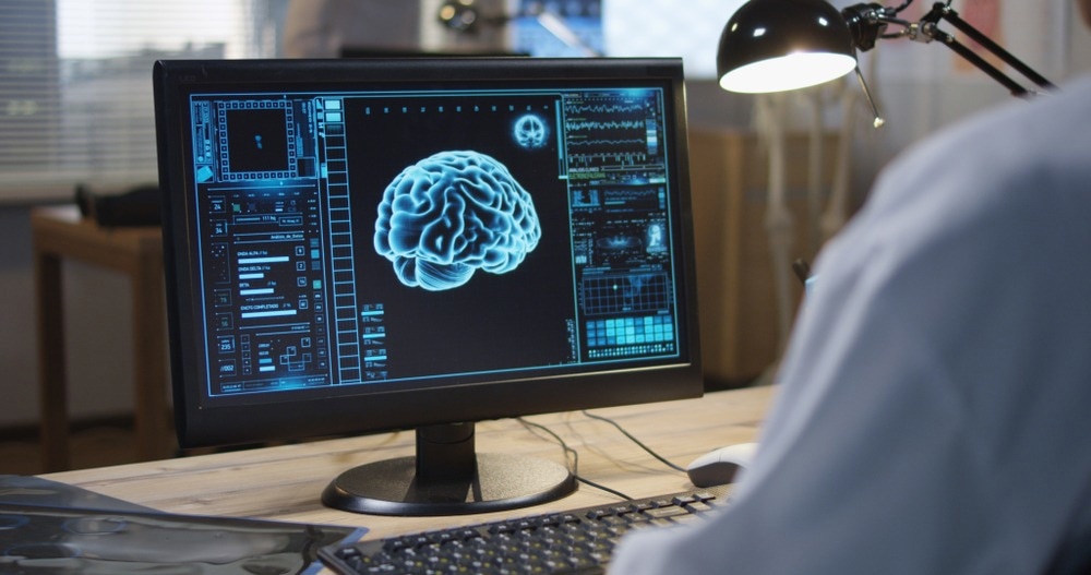
Neuroimaging. Image Credit: Frame Stock Footage/Shutterstock.com
Brain mapping at a cellular level: the connectome
Brain mapping can additionally be investigated at a cellular level and therefore gave rise to the study of the ‘connectome’, a “comprehensive structural description of the network of elements and connections forming the human brain”.
The Human Connectome Study aimed to characterize brain connectivity and function and their variability in healthy adults using various functional imaging modalities in combination with genetic and behavioral studies. With the aim of freely sharing tools and data, the Human Connectome Project has led to visually stunning maps of white matter tracts spanning throughout the brain, and ultimately leading to advancements in the emerging field of connectomics.
The use of this data by the University of Oxford led to the formulation of an axis between variation in brain connectivity and an individual’s traits. Using connectome data they identified that upon their axis classically positive behaviors demonstrated different connectivity to those with negative ones. Other publications have identified patterns of white matter connectivity that could be an important determinant of behavior and were associated with vocabulary compression and language performance.
Where will brain maps lead us in the future?
The future of brain mapping is likely to feature the collaborative efforts of functional neuroimaging, behavioral testing, electrophysiology and connectomics.
Brain mapping provides invaluable information regarding the localization of behaviors in neurology, and how perturbations can lead to cognitive and phycological conditions. Editing these maps may provide future personalized medicine for neurological disorders.
A mass of white matter tracts in the human brain. Data from The Human Connectome Study. www.humanconnectomeproject.org
References
- Breiter, H. C., Etcoff, N. L., Whalen, P. J., Kennedy, W. A., Rauch, S. L., Buckner, R. L., . . . Rosen, B. R. (1996). Response and Habituation of the Human Amygdala during Visual Processing of Facial Expression. Neuron, 17(5), 875-887. doi:https://doi.org/10.1016/S0896-6273(00)80219-6
- Cohen, J. D., Forman, S. D., Braver, T. S., Casey, B. J., Servan-Schreiber, D., & Noll, D. C. (1994). Activation of the prefrontal cortex in a nonspatial working memory task with functional MRI. Hum Brain Mapp, 1(4), 293-304. doi:10.1002/hbm.460010407
- Corbett, B. A., Carmean, V., Ravizza, S., Wendelken, C., Henry, M. L., Carter, C., & Rivera, S. M. (2009). A functional and structural study of emotion and face processing in children with autism. Psychiatry Res, 173(3), 196-205. doi:10.1016/j.pscychresns.2008.08.005
- Elam, J. S., Glasser, M. F., Harms, M. P., Sotiropoulos, S. N., Andersson, J. L. R., Burgess, G. C., . . . Van Essen, D. C. (2021). The Human Connectome Project: A retrospective. Neuroimage, 244, 118543. doi:https://doi.org/10.1016/j.neuroimage.2021.118543
- Raichle, M. E. (2009). A brief history of human brain mapping. Trends Neurosci, 32(2), 118-126. doi:10.1016/j.tins.2008.11.001
- Rombouts, S. A., van Swieten, J. C., Pijnenburg, Y. A., Goekoop, R., Barkhof, F., & Scheltens, P. (2003). Loss of frontal fMRI activation in early frontotemporal dementia compared to early AD. Neurology, 60(12), 1904-1908. doi:10.1212/01.wnl.0000069462.11741.ec
- Smith, S. M., Nichols, T. E., Vidaurre, D., Winkler, A. M., Behrens, T. E. J., Glasser, M. F., . . . Miller, K. L. (2015). A positive-negative mode of population covariation links brain connectivity, demographics and behavior. Nature Neuroscience, 18(11), 1565-1567. doi:10.1038/nn.4125
- Sporns, O., Tononi, G., & Kötter, R. (2005). The Human Connectome: A Structural Description of the Human Brain. PLOS Computational Biology, 1(4), e42. doi:10.1371/journal.pcbi.0010042
- Vaidya, A. R., Pujara, M. S., Petrides, M., Murray, E. A., & Fellows, L. K. (2019). Lesion Studies in Contemporary Neuroscience. Trends Cogn Sci, 23(8), 653-671. doi:10.1016/j.tics.2019.05.009
- Van Essen, D. C., Ugurbil, K., Auerbach, E., Barch, D., Behrens, T. E., Bucholz, R., . . . Yacoub, E. (2012). The Human Connectome Project: a data acquisition perspective. Neuroimage, 62(4), 2222-2231. doi:10.1016/j.neuroimage.2012.02.018
Further Reading
- All Neuroscience Content
- What is Neurology?
- What is the Difference between Neurology and Neuroscience?
- What is Neuroscience?
- What is Neurosurgery?
Last Updated: Jul 27, 2022

Written by
Olivia Grech
Olivia Grech is a Brain Research UK PhD student at the University of Birmingham studying mechanisms and new treatments for headache in raised intracranial pressure. Her project has also explored the metabolic effects of headache mechanisms in animal models in addition to analysing metabolic profile of patients with disorders of headache (Idiopathic Intracranial Hypertension). She has an MRes in Biomedical Science and Translational Medicine from the University of Liverpool and a BSc in Biomedical Sciences from the University of York.
Source: Read Full Article