
Vincent Groenhuis, a Ph.D. candidate at the University of Twente, has spent the past four years working on the fight against breast cancer. Using various prototypes of 3-D-printed biopsy robots, he has been able to develop methods which alleviate the suffering of patients and make breast cancer testing more accurate and efficient. Vincent Groenhuis was awarded his Ph.D. cum laude on 9 January. But for him, the work does not stop here.
Breast cancer is still the most common form of cancer in women. It is possible to obtain a tissue sample for testing by inserting a needle into the breast and manipulating it to the location of the abnormal tissue (lesion). Once further tests have been carried out, it is possible to arrive at an accurate diagnosis. This procedure is known as a biopsy.
Accurate manipulation of the biopsy needle is essential in order to fight breast cancer, as with all other types of cancer. By using a special needle, the tip of which can be heated to a high temperature (thermo-ablation) or cooled to a very low temperature (cryo-ablation), it is possible to destroy tumor cells close to the tip of the needle. In this way, the cancer can be treated without major surgical procedures.
Robotics is the solution
MRI scanners are capable of detecting and visualizing the location of abnormal tissue very accurately. However, when the biopsy needle is manipulated by hand, the results are sub-optimal. The solution lies in robotics, as Vincent has demonstrated once again in his dissertation, but this approach requires a great deal of expertise. “Not all robots can be used successfully in combination with MRI scanners. Robots are often made of metal and that can cause problems due to the strong magnetic fields created by MRI scanners,” he explains.
During his Ph.D. research, Vincent investigated various types of robot and produced a number of prototypes. He had previously worked on the Stormram and Sunram robots, of which there are now five. The newest version is the Sunram 5. MURAB is a different research line to Stormram and Sunram. There is only one finished MURAB robot. Both research lines have developed in parallel to one another, and each has its own philosophy. Vincent describes both projects in his dissertation.
MURAB
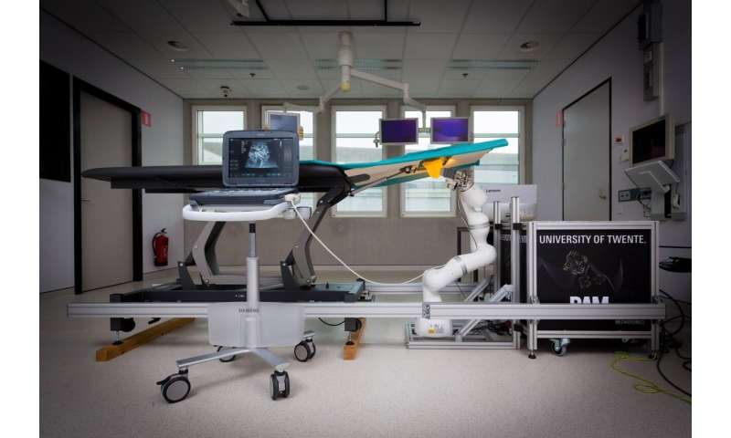
MURAB, which stands for MRI And ultrasound robotic assisted biopsy, is a robot outside the MRI scanner that combines ultrasound, MRI, elastography and computer vision to implement the correct biopsy strategy based on a patient-specific model. The robot takes deformations into account. For more information, watch the video below.
Stormram
The predecessor to this robot, Stormram 4, is made of plastic by a 3-D printer and is powered by pressurized air. The advantage of plastic is that the robot can be used inside an MRI scanner. Previously, the robot won a prestigious prize at the Surgical Robotic Challenge at the international Hamlyn Symposium in London. This is one of the world’s most important events in the field of robotic surgery.
The Stormram has brought new energy to breast cancer diagnostics. This is due to its highly accurate use of the needle, virtually real-time scanning using MRI and a single thin needle biopsy, all of which makes diagnosis faster and more accurate.
The Stormram is driven by straight and curved hydraulic motors, and the robot is controlled using 5-meter long hoses outside the MRI scanner. The design is smaller than the previous version, meaning that it can fit into the narrow tunnel of the MRI scanner.
The future
Source: Read Full Article