Introduction
Functional magnetic resonance imaging (fMRI)
Functional neuroimaging in the monitoring of sleep patterns
Using positron emission tomography (PET) to treat lymphoma
References
Further reading
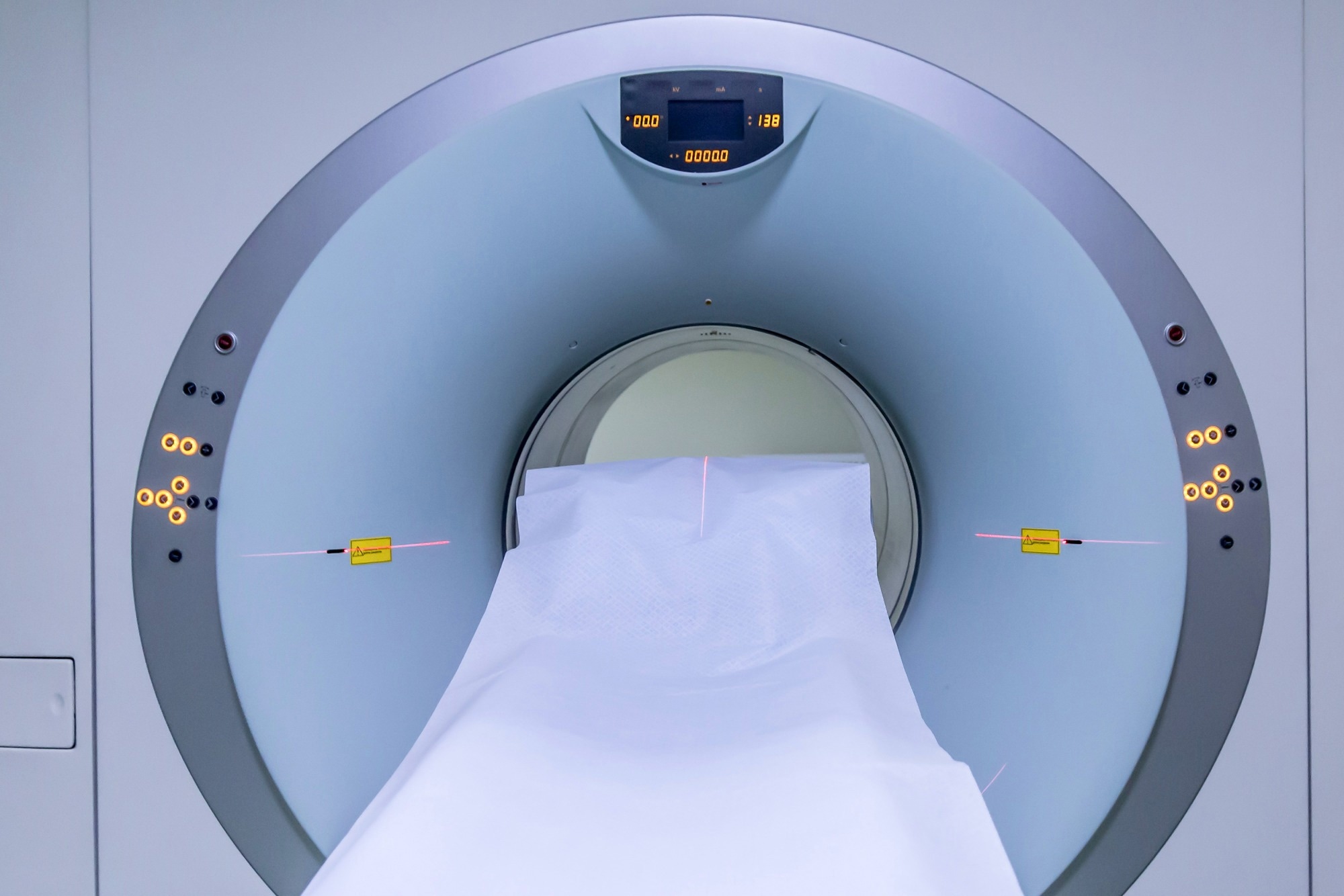
Most people would categorize computerized tomography (CAT) scans, positron emission tomography (PET) scans, and magnetic resonance imaging (MRI) scans into the same grouping. However, it is important to note that significant variability exists between these neuroimaging techniques. Often separated into two sets, there exists structural neuroimaging and functional neuroimaging.
While structural neuroimaging is used to segregate and assay different brain regions, like the shape of grey matter, cerebrospinal fluid, et cetera., functional neuroimaging incorporates the measure of neural activity. This assay of neural activity, measured through electrodes, magnetic and radio waves, and X-Rays, is used to assess mental function.

Image Credit: Said Marroun/Shutterstock
Functional magnetic resonance imaging (fMRI)
Often performed after a diagnosis is achieved, fMRI’s serve mainly to see whether surgery is a favorable option or not. Therefore, scans of this nature are performed roughly 24-48 hours before the scheduled surgery. The images help surgeons navigate through the human body, while in the operating room, showing the points of entry, the location of masses, and other important information. This differs from regular MRIs, which assess other patients' organs, bones, or tissues.
fMRIs, and MRIs in general, use the duality between magnetic fields and radiofrequency energy to scan the brain. In actuality, the X-rays portray the hydrogen atoms responding to these pulses in energy. This apparatus uses the magnetic properties of living tissue to provide the contrast needed to generate these images. The energy emitted during the relaxation time (T1) of these hydrogen atoms is what the MRI scanner will pick up while the computer reconstructs this information to provide us with an image. If tumors or other major entities are present amidst the brain matter, the use of contrast dye will sometime be employed. This enhances the clarity and visibility of whatever the dye meets, such as target tissues or blood vessels.
A common mistake amongst aspiring medical students and radiologists is that computerized axial tomography (CAT) scans are a type of functional neuroimaging. Conversely, these series of X-ray images combine to reveal the size and shape of structures in the brain, falling under the practice of structural neuroimaging. Amongst other limitations, CAT scans do not have a high enough spatial resolution to be considered functional neuroimaging.

 Read Next: Should MRI be Used to Diagnose Prostate Cancer?
Functional neuroimaging in the monitoring of sleep patterns
Sleep is now being monitored in research settings using electroencephalography (EEG), a methodology that has been in use for a long time, speaking volumes about its practicality. The digital graph resulting from the information provided by electrodes will portray time on the x-axis and frequency in Hz on the y-axis. This will effectively monitor “brain waves,” also known as neural oscillations. The frequency of these brain waves is segmented into bands, each relating to different brain functions. There are oscillations produced at the delta band (0.5-4Hz), which are linked with deep sleep, theta waves (4-8Hz) associated with meditation, and alpha waves (8-12Hz), which indicate an extremely mild awareness. Though we are still aloof about which band corresponds to which specific action, it has been established that the higher frequency these waves are, the more alert the individual will be.
By studying the interworking of these oscillations during hours of sleep and activity, we garner essential information about brain health. Abnormal brain health, and an irregularity in circadian rhythms, have exhibited the amplification of Alzheimer’s symptoms and greater susceptibility to prion diseases. Through the use of EEG, we can monitor neuronal oscillations after some treatment or change of lifestyle is expressed, granting us a better idea of what behavior or treatment must be undergone. In addition, we see how much sleep an individual truly needs during a specified age in their life.
Using positron emission tomography (PET) to treat lymphoma
PET scans, another imperative form of functional neuroimaging, are accomplished by first injecting the patient with a radiopharmaceutical-like simple sugar, most often referred to as fluorodeoxyglucose (FDG). The apparatus will then pick up the gamma rays as the sugar flows through the bloodstream. Surveying this blood flow through the brain will highlight regions of the brain where more activity is present, useful when diagnosing aphasia, brain lesions, irregular masses, and other brain disorders.
While these scans are not ideal for all variants of cancer, it is essential in the monitoring of Hodgkin’s lymphoma, Burkitt lymphoma, and other T-cell lymphomas. PET can detect sites of disease spread within small nodes that would otherwise be undetectable using a CT scan or other form of X-ray technology. PET can also monitor within the bone marrow, a feature that CT scans do not possess. To know where cancerous masses are and how far they have metastasized is to know how to treat the disease. By determining what stage of cancer one has, we are made aware of how many cycles of chemotherapy and radiotherapy are required and in which areas they should be administered.
Before the normalization of PET scans, bone marrow biopsies were common practice – a procedure that could cause extreme pain, bleeding, infection, and death. This technique, amongst other function neuroimaging techniques, has greatly contributed to healthcare and medicine.

PET can be used to treat lymphoma. Image Credit: Lightspring/Shutterstock
References
- Klimesch W. (2012). α-band oscillations, attention, and controlled access to stored information. Trends in cognitive sciences, 16(12), 606–617. https://doi.org/10.1016/j.tics.2012.10.007
- Khanna, N., Altmeyer, W., Zhuo, J., & Steven, A. (2015). Functional Neuroimaging: Fundamental Principles and Clinical Applications. The neuroradiology journal, 28(2), 87–96. https://doi.org/10.1177/1971400915576311
- Chow, M. S., Wu, S. L., Webb, S. E., Gluskin, K., & Yew, D. T. (2017). Functional magnetic resonance imaging and the brain: A brief review. World journal of radiology, 9(1), 5–9. https://doi.org/10.4329/wjr.v9.i1.5
- Gallamini A, Borra A. Role of PET in lymphoma. Curr Treat Options Oncol. 2014 Jun;15(2):248-61. doi: 10.1007/s11864-014-0278-4. PMID: 24619427.
- Dang-Vu, T. T., Schabus, M., Desseilles, M., Sterpenich, V., Bonjean, M., & Maquet, P. (2010). Functional neuroimaging insights into the physiology of human sleep. Sleep, 33(12), 1589–1603. https://doi.org/10.1093/sleep/33.12.1589
- Nofzinger EA. Functional neuroimaging of sleep. Semin Neurol. 2005 Mar;25(1):9-18. doi: 10.1055/s-2005-867070. PMID: 15798933.
Further Reading
- All Neurology Content
- What is Neurology?
- What is the Difference between Neurology and Neuroscience?
- What is Neuroscience?
- What is Neurosurgery?
Last Updated: Sep 28, 2022

Written by
Vasco Medeiros
Obtaining an International Baccalaureate Degree at Oeiras International School, with higher levels in Chemistry, Biology, and Portuguese, Vasco Medeiros has just graduated from the University of Providence College with a Bachelor of Science. Before his work as an undergraduate, he first began his vocational training at the HIKMA Pharmaceuticals PLC plant in Ribeiro Novo. Here he worked as a validation specialist, tasked with monitoring the gauging and pressure equipment of the plant, as well as the inspection of weights and products.
Source: Read Full Article
